
By trauma, infection and
Operation
Segmental defects of the mandible caused by tumor resection are not only related to the maintenance of facial contour and shape, but also seriously affect the patient
Oral cavity
Chewing, speech and swallowing functions.The reconstruction of mandibular defect has become the oral and maxillofacial
surgical
I have been paying attention during the operation
clinical
problem. The repair of mandibular defect not only needs to restore its physiological function, but also its anatomical shape and aesthetic effect should be considered.
For repairing mandibular defects
Implants
Design, the traditional shape-oriented bionic design has gradually been transformed into a functional bionic design. Taking into account the individual differences and the complexity of the disease, custom design is advocated to promote the restoration of function and aesthetics. In addition, with the advancement of additive manufacturing technology,3D printingProvided technical support for the manufacture of custom implants and caused
biology
medicine
Great interest in application.
The purpose of this research is to develop a3D printingA new functional design method of PEEK implants and fibular autografts for severe mandibular defects.
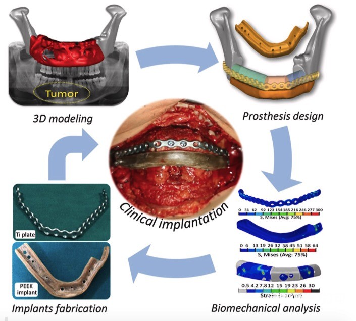
The CT scan of a 39-year-old man showed a 71mm×31mm×22mm tumor on the right mandible, using Mimics
software
Construct a 3D model of the mandible and tumor from the CT data, and perform simulated resection of the tumor and the corresponding mandibular segment.In meeting the three designs of safety, functionality and shape consistency
standard
In the case of fibula autograft and3D printingThe combined method of PEEK implants repairs mandibular defects.
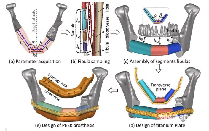
The design workflow is shown in Figure 2. First, the key feature parameters (LR, LM, LL, α, β) are collected in the defect area to guide the subsequent fibula sampling.Secondly, based on the above parameters, generate fibula and
Blood vessel
3D model, and determine the spatial position according to the occlusal datum and three height parameters (hR, hM, hL). Third, the fixation of the fibula segment is completed by designing a fixation plate that perfectly matches the fibula. Finally, use Boolean operations to establish the macroscopic shape of the PEEK implant on the original mandible and fibula, and fix the prosthesis around the mandible and fibula with screws.
Through finite element analysis, the biomechanical behavior and biomechanical characteristics of the jaw reconstruction of various masticatory movements were studied. The results showed that the maximum Von Mises stress of each component was lower than the yield strength of the corresponding material, and the safety factor was greater than 2.3 times, indicating that the restoration can be guaranteed The safety of the method. Finally, this new repair method was applied to the clinic and achieved good clinical results.
references:
Kang J, Zhang J, Zheng J, Wang L, Li D, Liu S. 3D-printed PEEK implant for mandibular defects repair-a new method. J Mech Behav Biomed Mater. 2021;116:104335.
(Editor in charge: admin)
0 Comments for “3D printed PEEK implants to repair mandibular defects-a new method”