Guide:
existbiologyIn the field of printing, researchers have designed a new six-degree-of-freedom robot arm that cooperates with biological3D printingfunction, constructing beating myocardial tissue, these in vitro structured myocardial tissue have been successfully beating continuously for 6 months
.
 .
.
26 February 2022 It was learned that researchers from the Chinese Academy of Sciences have managed to overcome the traditional3D printingThe method combines the difficulties of creating artificial heart tissue with a 6-degree-of-freedom robotic arm to create a viable myocardial tissue.This novel 3D printing method not only succeeded in making a viable
Blood vessel
Organoids, and remained alive Mars for 6 months. The paper is titled “A Robot-Based Multiaxial Bioprinting System to Support Natural Cell Function Preservation and Cardiac Tissue Fabrication”, and the abstract of the paper is as follows:

Although artificial tissue and organ engineering have made great progress in recent years,
But how to generate large-scale, functionally complex organs in vitro is still a matter of regenerationmedicinea big challenge
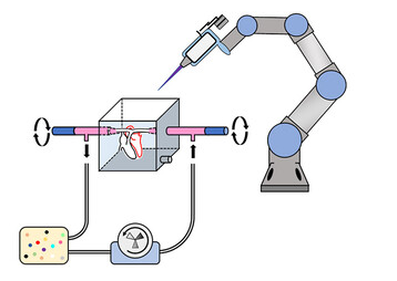
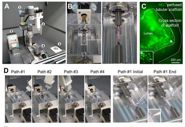
. 3D bioprinting has demonstrated its ability and advantages to create simple tissues, but it remains difficult to generate vasculature and maintain cellular function in complex organ production. Here, we overcome the limitations of conventional bioprinting systems by incorporating a six-degree-of-freedom robotic arm with a bioprinter, enabling cell printing on 3D complex-shaped vascular scaffolds from all directions. We also developed an oil bath-based cell printing method to better preserve the natural function of the cells after printing. Combined with a self-designed bioreactor culture strategy, our bioprinting system is able to spontaneously generate blood vessels and form contractile and long-term viable cardiac tissue.
This bioprinting strategy mimics the developmental process of in vivo organs and provides a promising solution for the in vitro fabrication of complex organs.

Typical 3D layered printing methods are not suitable for creating complex vascular networks because stacking often damages cells, and the biomaterials that bind them together prevent them from functioning as organ tissues.
That’s why the researchers programmed the robotic arm with six rotating joints to seed cells on the scaffold from all directions, in a process that does little damage, gradually generating endothelial cells until new capillaries form. Not only that,
They also used two of these robotic arms to simultaneously seed different cell types, such as cardiomyocytes, on the scaffold, a major advance in the tissue engineering community
.

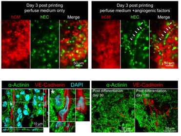
By applying this printing and differentiation strategy, the researchers finally fabricated a piece of vascularized myocardial tissue (2 cm in length, 200-500 μm in thickness, and ∼1.256 cm in total area) on a tubular scaffold that remained contracted for at least 6 months. We performed histological analysis of cardiac tissue on days 30 and 180 in vitro and found no significant tissue damage at either time point. In addition, cardiomyocytes exhibited a good Z-stripe arrangement, indicating that the fabricated cardiac tissue had developed and maintained an intact myofibrillar structure, which is the physiological basis for cardiac contraction.

(responsible editor: admin)


0 Comments for “A new 6-degree-of-freedom robotic arm that cooperates with 3D printing to create beating myocardial tissue”