
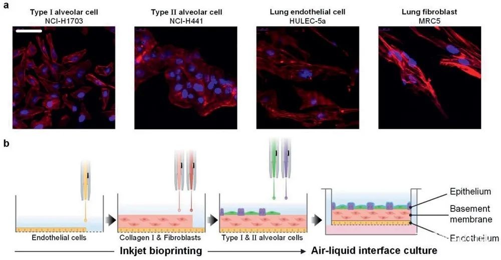
Respiratory diseases seriously endanger people’s health, and there is an urgent need for related physiological models of the human respiratory system to study the pathogenesis, drug efficacy and pharmacy of the disease.The Sungjune Jung team and Hwa-Rim Lee team from Pohang University of Science and Technology in South Korea used cell-loaded inkjet printing technology to print alveolar models with four human alveolar lines, namely type I and type II alveolar cells (NCI-H1703 and NCI- H441), lung fibroblasts (MRC5) and lung micro
Blood vessel
Endothelial cells (HULEC-5a). This model can replace traditional pathology and pharmacology testing models to a certain extent. The related paper “All-Inkjet-Printed 3D Alveolar Barrier Model with Physiologically Relevant Microarchitecture” was published in the journal Advanced Science.
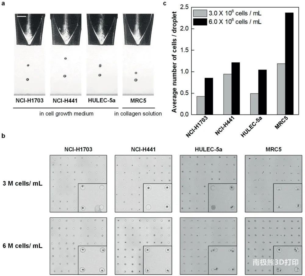
First, the research team printed an ultra-thin three-layer alveolar model by layer-by-layer cell-loaded inkjet printing technology, and optimized the inkjet printing process to determine the concentration of each cell ink to produce a high-resolution and Accurate 3D alveolar model.
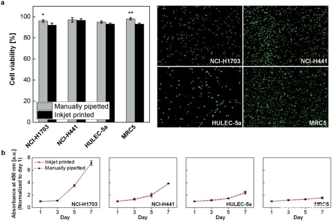
Next, the research team evaluated the cell viability of inkjet printing. During the experiment, no significant cell death was observed.
biology
Printing will not adversely affect alveolar cells.
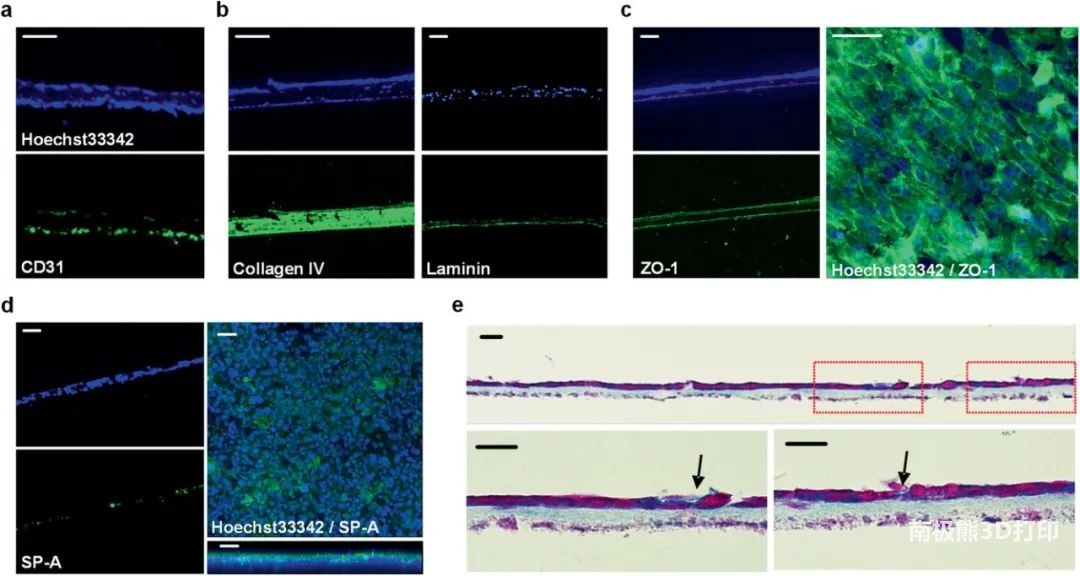
Subsequently, the research team evaluated the structural characteristics and barrier properties of the alveolar model. The three-layer structure can be clearly seen through histological images, with epithelial and endothelial cells evenly distributed on both sides of the collagen basement membrane. The structural composition and function of the alveolar barrier were studied by immunohistochemical analysis.
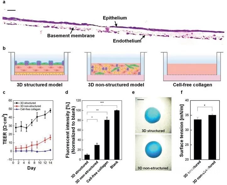
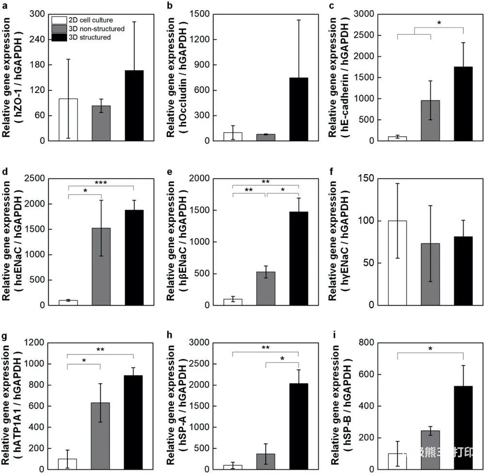
Immediately afterwards, the research team conducted a qPCR test to quantify the level of representative genes and used it to study cell function in the alveolar model. The results confirm that the 3D structural model has tighter barrier properties compared to the 2D model.
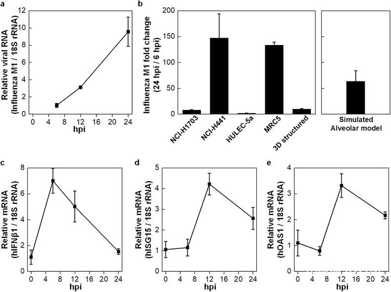
Finally, the research team used H1N1 influenza A virus (PR8 strain) to act on the air side of the alveolar barrier model to simulate influenza infection in the epithelial layer of the respiratory tract. The results showed that the infection rate of NCI-H1703 and HULEC-5a cells at 24 hours was 7.7 times and 1.5 times that of 6 hours.
In short, the research team used high-resolution inkjet bioprinting technology to reconstruct four basic alveolar models of alveolar cells in a 3D multilayer structure. It can precisely control the spatial arrangement and population of multiple cell types layer by layer, and reproduce the complex microstructure and morphology of the alveoli and the main functions of the alveoli, such as barrier integrity and surfactant secretion. Although the model is a three-dimensional structure, it is a flat shape and cannot imitate the 5% linear elongation change of physiologically inhaled air. If this structural and functional problem is overcome in future work, inkjet-printed in vitro 3D alveolar barrier models may become a new platform for studying respiratory diseases and drug efficacy.
(Editor in charge: admin)
0 Comments for “Advanced Science: Inkjet printing 3D alveolar model”