
China3D printingNet April 16th, in the final degree program of Biomedical Engineering at the Polytechnic University of Catalunya, Antonio Molina Herrero (Antonio Molina Herrero) solved the challenge of bone regeneration in bioprinting.Through research3D printingAnd various material forms, this student researcher recently published ” 3D printingHis findings are described in detail in the structure characteristics of calcium phosphate obtained.
When analyzing the geometry of filaments, the porosity in the structure, and properties such as surfaces and concave surfaces, Herrero used microtomography technology to compare with other images, calculations, and measurements, and finally allowed researchers to compare nozzles and printing There is a better understanding of the deviation between materials and how to better use calcium phosphate to construct scaffolds for bone regeneration in tissue engineering.
Among synthetic bone grafts, bone grafts based on calcium phosphates (CaPs) have been extensively studied. However, they cannot sufficiently promote osteogenesis.
The interconnected pore network can vascularize all areas and colonize the tissues later.Many studies have been conducted to find the best pore size in the stent[1], But the conclusion is not clear, and some studies contradict other studies. It is clear that the hole must be large enough to allow vascularization (50 μm), but if the hole is larger than a certain size (500 μm), the stent will no longer act as a stent. “
Herrero (Herero) emphasized the key to his discovery of the research bracket: the shape of the hole. Explained that many methods have been used for CaP porous structures, such as granulation, foaming, leaching and freeze-drying. With the advent of direct ink writing (DIW), this structure has greater potential for success; however, in vivo testing and further Characterization still faces challenges.

Schematic diagram of custom modular nozzle design. Red: The top of the nozzle has a standard “Luer lock” connector on the top. Gray: The lower part of the nozzle. It cannot be screwed in to fit a custom disk. Turquoise: A disc with a variety of center hole designs.
“The complex and corrugated shapes of these 3D structures can be studied through advanced imaging techniques, such as Micro-CT and Scanning Electron Microscopy (SEM). At the same time, the image processing that extracts the quantitative information of these images is Promising and challenging. These tools are being used more and more because of their immediacy and reliability. With them, it is possible to study the morphological characteristics of three-dimensional structures, and to design and design new scaffold shapes for tissue engineering. Characterization provides interesting morphological and structural information.”
Prepare and check samples, and compare images and calculation results.
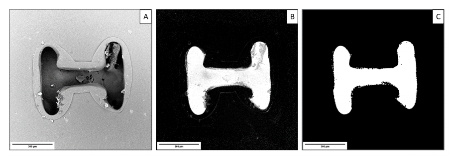
Mode 1 image segmentation process: A) original original image, B) image segmented with Ilastik and C) image after Fiji noise reduction. 300μm scale bar
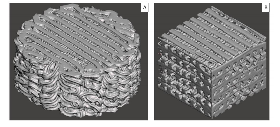
Mode 2 applies VOI clipping before (A) and after (B).
Four technologies were evaluated:
.
computer
.image
.Grid Mixer
.Manual practice
Five sample patterns were evaluated and numbered from control number to 1-5.

SEM nozzle image; right: sagittal view of Micro-CT scaffold.
“In the porosity results, there is not much difference between the scaffolds. This is because the scaffolds are printed under preset conditions, so they will have the same porosity, and the samples between them (250μm) can be compared. Pore size. The distribution result is a good indicator. The peaks in the figure represent regularity and good print quality.” Herrero concluded.
In the specific surface area, the pattern with the highest coefficient is 5, while the control group is the lowest. Since the control part is round, these results are coherent, so it has the smallest surface/area coefficient and pattern 5; although small, it repeats very frequently, resulting in the sum of all filaments having a larger surface than other patterns. In addition, the MeshMixer method has too many errors compared with other methods. This may be due to how the plug-in 3D Viewer performs the meshing process from the Micro-CT bracket before the meshing. However, more experiments are needed to ensure this. A hypothesis. “
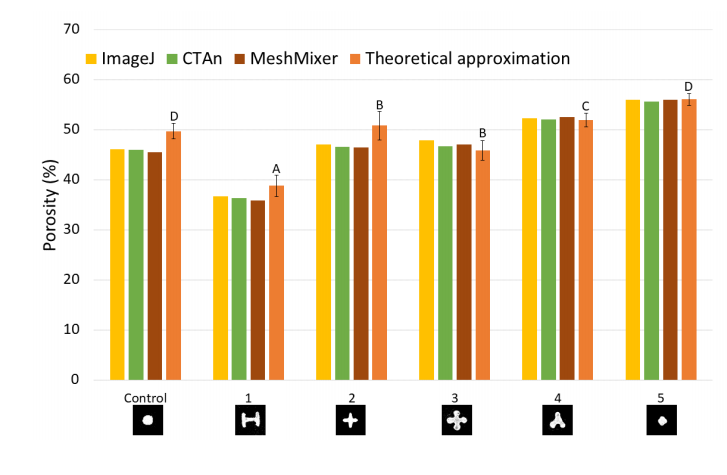
Percentage of porosity. Statistically significant differences indicated by different letters (p = 0.05)
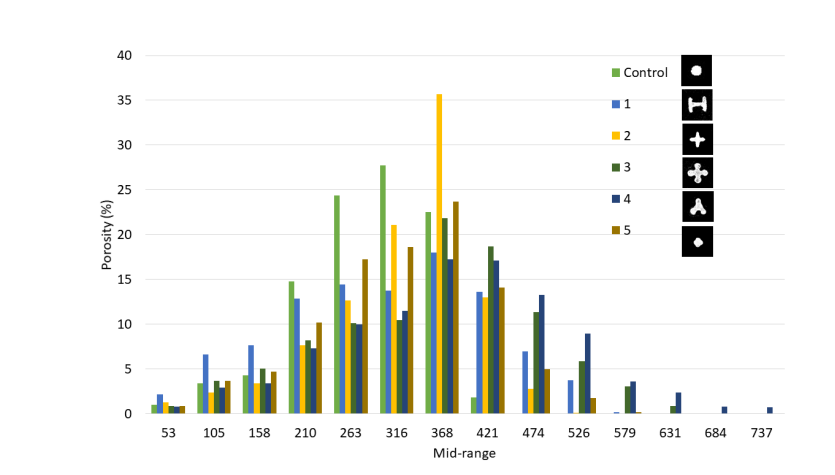
Pore size distribution.
China3D printingOnline reviews: The research of bone regeneration scaffold is underway and has participated in a series of projects and experiments. These researches and experiments use materials such as PLA, coated nanofibers and bio-printing and dyeing with antibacterial properties.
(Editor in charge: admin)
0 Comments for “Bone Regeneration Scaffold: Using 3D Printing to Study the Structure of Calcium Phosphate”