
China3D printingNet, June 24, researchers from Teeside University in the United Kingdom have used3D printingAnd scanning technology advances a key aspect of forensic investigation: physical fitness analysis (PFA). Using and comparing two different 3D imaging methods, the research team was able to reproduce human bone fragments for use in the PFA process.This not only prevents unnecessary damage to real evidence during a crime scene investigation (CSI), but it also extends3D printingLegal application.
“Fused Filament Manufacturing (FFF)3D printingProven to be an accurate and useful method for creating physical replicas of bone fragments, performing physical fit analysis (PFA) and bone fragment reconstruction. Therefore, we recommend combining μCT imaging with FFF 3D printingUsed in conjunction, this is an excellent choice for non-destructive physical fitting confirmation when dealing with small fragments and burned bones. “The research team said.
To evaluate the best of PFA reconstruction3D scanningIn the process, the researchers produced two sets of bone fragments (pictured). The picture comes from Science Direct.
Analysis of physical fitness in crime scene investigation
CSI usually requires investigators to examine a series of objects as evidence, including human remains, some of which may be damaged or broken due to trauma suffered during the event. These remains are usually determined by PFA to determine their suitability. If this process leads to good physical adaptation, it may place the suspect at the crime scene or facilitate the reconstruction of objects that may solve the case. However, PFA involves a lot of matching and manual handling matters, which may cause fragments to be damaged during the handling process.
In addition, in some cases, PFA can be extremely challenging, such as fragments that may constitute a biohazard, are very small, or the bones themselves are too fragile to move. Considering that the reconstruction process usually involves gluing parts together, this can cause problems and prevent researchers from fully understanding the nature of the trauma. This makes certain shapes of bones difficult to record or present, especially those fragments with complex three-dimensional properties or fragments embedded in external materials. As a result, this situation is obviously not enough to make a statement in court.
although3D scanningAnd modeling has been used in a series of forensic anthropology applications, but the processing and reconstruction of bone fragments is still a problem. For example, 3D modeling is currently used in dismemberment, weapon matching, skull measurement and facial reconstruction cases.Scanning allows to obtain high-resolution images at the nanometer level in some cases, but they are oftenTime consumingIt is expensive and requires professional knowledge and software to operate. On the other hand, surface scanning methods tend to be cheaper and easier to use, and are often used for post-mortem quantitative damage analysis, marking and soft tissue damage analysis.
At the same time, additive manufacturing has proven to be accurate enough to produce dental models for maxillofacial surgery.The research also proved3D printingIn the visualization and analysis of forensic evidence, researchers set out to combine these techniques to create3D printingReplica of skull fracture. These 3D models provide the possibility of PFA occurrence without having to deal with the original evidence fragments too much, while minimizing the risk of damage or contamination. In addition, such models provide 360-degree visualization in a compelling and easy-to-understand format and can be used to improve jury comprehension during trials.

3D printing
The copy, compared with the SLS scan model (left), the μCT scan model (right) shows stronger ridge details” alt=” of the virtual model
3D printing
The copy, compared with the SLS scan model (left), the μCT scan model (right) shows stronger ridge details” width=”620″ height=”237″ />
Virtual model3D printingIn the copy, the μCT scan model (right) shows stronger ridge details compared to the SLS scan model (left). The picture comes from Science Direct.
3D printingSkull fragment copy
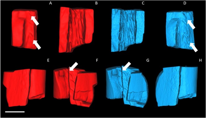
The researchers compared the volumetric scanning technique micro-computed tomography (μCT) with the surface scanning technique structured light scanning (SLS) to assess the advantages and disadvantages of the two methods.To test its potential for PFA, the team3D printingTwo burn bone fragment models were created to simulate the damage that may be encountered in a real investigation.
Using the archeological human femur donated by the University of Portsmouth as a model, the replicated bone sample was cut and burned in a Gallenkamp muffle furnace at 600°C for 30-60 minutes. During the combustion process or cooling process, each part of the bone will naturally split into at least two separate fragments longitudinally. Perform 3D imaging and printing of two adjacent fragments to evaluate their techniques for visualizing and analyzing the physical suitability of burned bone fragments.
The SLS scanner used for testing was Shining 3D EinScan Pro +, and the larger μCT Zeiss Xradia 520 Versa scanner was also selected for its simple setup and non-destructive process.After scanning, use PLA filament to scan the fragments with FFF Prusa i3 desktop printer3D printing. Choose the best print quality (0.15 mm) and set the fill amount to 0% to create a completely hollow print.Then, in pairs3D printingPFA is performed on the skeletal model, and its accuracy depends on the feature matching and alignment between the two fragments and the tactile “feel” of the coordination. 3D printingOf femur fragments (as shown in the picture) can be put together again” alt=”two sets of burnt3D printingThe femur fragments (as shown in the picture) can be put together again” width=”620″ height=”301″ />
Two sets of burnt3D printingThe femur fragments (as shown in the picture) can all be put together again. The picture comes from Science Direct.
Find3D printingA high level of detail is retained in both the μCT and SLS models. In general, the resulting print quality is sufficient to perform PFA. According to the “fit quality” standard set by the team, it was found that compared with SLS printing, the use of μCT printing can more easily confirm whether the body is suitable. In addition, in all the segment pairs created, the μCT model provides a tighter and firmer fit, resulting in a more detailed surface structure, which is of great significance in feature matching.
China3D printingOnline reviews: Can use FFF 3D printingTo produce copies of bone fragments with a sufficient level of detail, so that any kind of3D scanningTechnology. In addition, the team recommends selective laser sintering (SLS) by eliminating the need for support structures used in the FFF production process3D printingIt can prove to be a more effective way to produce models in future research. The research team said that although it turns out to be more expensive to implement, SLS printing will also bring a higher surface finish.In addition, the bone fragments successfully replicated can be used for other aspects of the PFA process.3D printingOpen a new application.
Compared with traditional methods, the application of 3D imaging and printing to PFA has many advantages. The virtual reconstruction of highly fragmented, fragile and possibly embedded remains provides an opportunity for complete reconstruction without damaging the original bone fragments.In addition, it can be scaled up to scale up from very small fragments or bones3D printingParts, generate 3D replicas to visualize the assembly and perform PFA on previously challenging projects.
China3D printingnetworkCompile the article!
(Editor in charge: admin)
0 Comments for “British researchers use FFF 3D printing to improve CSI analysis of skull fragments”