
On August 23, 2021, scientists from Tel Aviv University succeeded in 3D
biology
The brain tumor model was printed, forming the most complete laboratory growth model to date.
They printed a glioblastoma in a brain-like environment, including those that provided blood for the lump
Blood vessel
. Researchers say this is the most complete replication of tumors and surrounding tissues to date, and this breakthrough may help develop treatments. Currently, this research paper is published in “
Science Advances
“superior.
Glioblastoma is rare, but it is particularly scary. It grows rapidly on the brain or brainstem, cannot be cured, and is almost fatal.
Because this cancer is very aggressive, treatment usually requires chemotherapy and radiotherapy, and patients are often unable to complete these treatments because of their serious illness.
Glioblastoma tissue is extracted and cultured from tumors removed from patients. Doctors hope to use this method to further study this cancer. Ronit Satchi-Fainaro, a cancer researcher and nanoscientist at Tel Aviv University, said: “This is usually done in a petri dish. This is a very useful tool, but this approach has limitations.”
In a previous study, she and her team discovered a protein called P-Selectin, when glioblastoma cancer cells encounter microglia in the brain (the most prominent in the central nervous system) Immune cells).
This protein triggers microglia to take action to support glioblastoma, rather than fight it-a devastating result for people.
“we are at
Operation
This protein was found in tumors resected in China, but not in glioblasts grown on two-dimensional plastic petri dishes in our laboratory. “
“The reason is that cancer, like all tissues, behaves very differently on plastic surfaces than in the human body. About 90% of experimental drugs are
clinical
The stage failed because the success achieved in the laboratory cannot be reproduced in the patient. “
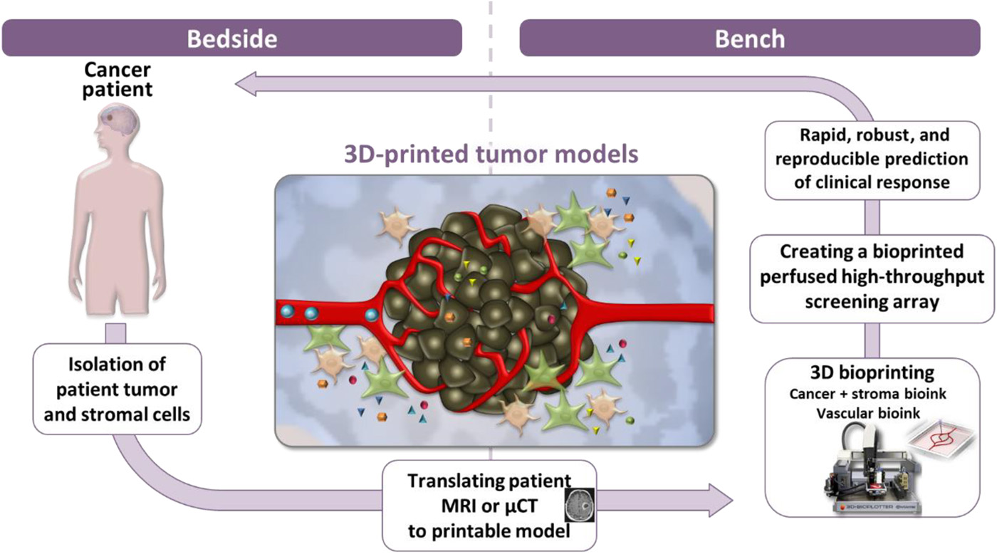
Therefore, the team of scientists trying to find a way to solve this limitation is glioblastoma bio-ink, which is created by glioblastoma cells, astrocytes and microglia from patients. Using removable bio-ink coated with cell types that form blood vessels, they also provide a functional blood supply for the model.
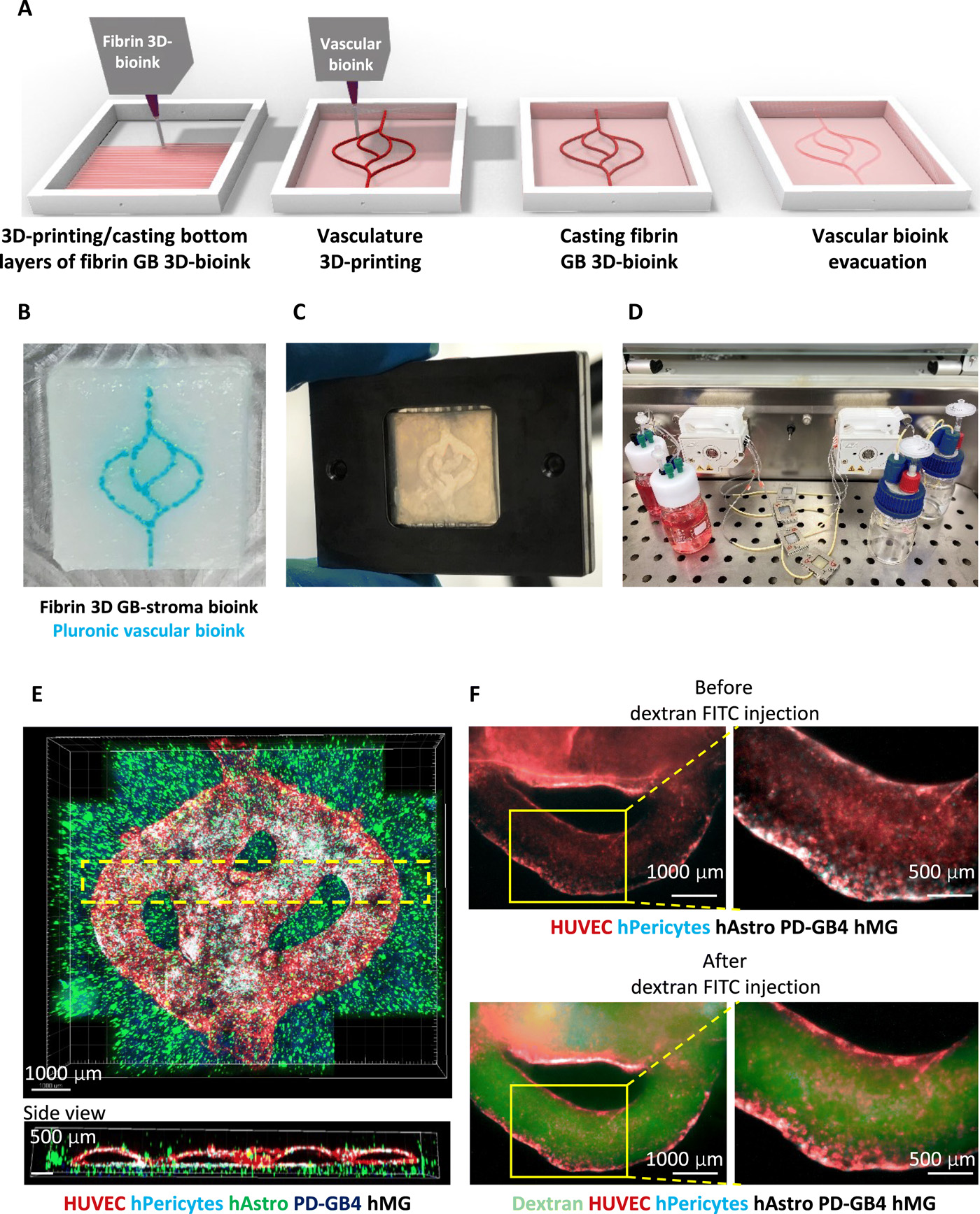
Each glioblastoma model is in a bioreactor3D printingComing out, this bioreactor is based on a hydrogel of extracellular matrix from the patient.
Satchi-Fainaro said: “The physical and mechanical properties of the brain are different from other organs, such as skin, breasts or bones. Breast tissue is mainly composed of fat, and bone tissue is mainly calcium; each tissue has its own characteristics, which affect The behavior of cancer cells and their response to drugs. Planting all types of cancer on the same plastic surface is not the best simulation of the clinical environment.”
The team then tested their model with P-Selectin.Glioblastoma cultures grown in petri dishes and3D printingA P-Selectin inhibitor was introduced in the model and animal model of. In the petri dishes, no changes were observed in growth or cell migration compared to the untreated control group.
for3D printingIn models and animal models, the P-Selectin inhibitor resulted in a slower growth rate compared to the untreated control group.
Saitchi-Fainaro said: “This experiment shows us why potentially effective drugs rarely enter the clinic, simply because they fail the test in the two-dimensional model, and vice versa: why it is considered an amazing success in the laboratory The drug eventually failed in clinical trials.
(Editor in charge: admin)
0 Comments for “Complete tumors are 3D printed to facilitate faster treatment”