
In the study of myocardial contraction in vitro, the layered micro-architecture that mimics myocardium in vitro plays an important role in cardiac tissue engineering.Recently, the research team of Xi’an Jiaotong University used molten electrofluid dynamics3D printingTechnology, printed a multi-scale, multi-directional fiber conductive scaffold to simulate heart tissue, thereby guiding the synchronous contraction of cardiomyocytes.
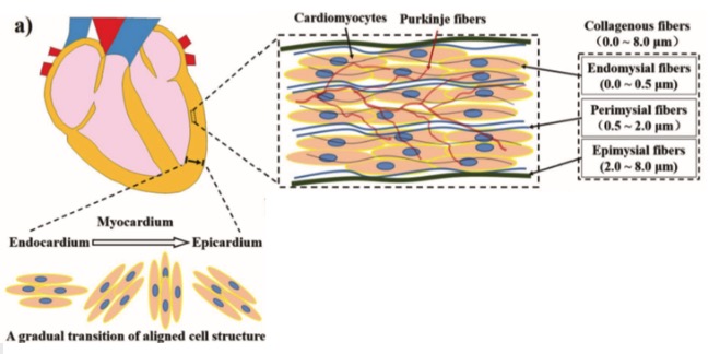
Cardiomyocytes are different from other cells. Cardiomyocytes beat autonomously, rhythmically, and contract synchronously. Based on this, the structure of the myocardium also has a certain particularity. From the inner membrane to the outer membrane, the myocardium is composed of multiple layers of electrically active tissues with specific orientations. , And distributed a variety of fibers. These fibers guide the growth and contraction of cardiomyocytes, and Purkinje fibers with good conductivity play an important role in the synchronous contraction of cardiomyocytes.
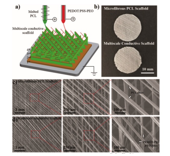
The research team of Xi’an Jiaotong University used electrohydrodynamics technology to print out micron-sized PCL fibers that simulate collagen fibers with polycaprolactone, and printed out the simulated conductive Purkinje fibers with polyethylene oxide and PEDOT:PSS solution. Nano-level PEDOT: PSS-PEO conductive fiber. Arrange the PCL fibers and conductive fibers in parallel with a certain direction interval, rotate 45 degrees and then lay another layer, and so on, lay four layers, thus forming a multi-layer oriented conductive scaffold to simulate the structure of the myocardium with a specific orientation. . Planting cardiomyocytes on this multi-scale conductive scaffold can make cardiomyocytes grow according to the direction of fiber arrangement and enhance the communication between cells to achieve synchronous contraction.
Electrofluid dynamics3D printingTechnology provides an innovative method for special muscle tissues to meet the different needs of specific tissues. This multi-scale conductive scaffold can better guide the specific arrangement of cell layers than single-scale scaffolds, and provides innovations for the in vitro culture of cardiomyocytes. Ideas.
The use of the multi-scale scaffold in this experiment to culture cardiomyocytes has an important role in the study of cardiomyocytes, and also provides the possibility of heart regeneration, and is of great significance to the treatment of heart-related diseases.
references:
Qi Lei, Jiankang He and Dichen Li, Electrohydrodynamic 3D printing of layer_specifically oriented, multiscale conductive scaffolds for cardiac tissue engineering, Nanoscale, 2019,11, 15195-15205.
(Editor in charge: admin)
0 Comments for “Electrohydrodynamic 3D printing of multi-scale conductive scaffolds for cardiac tissue engineering”