
China3D printingNet July 16th, a group of researchers from the University of Tampere in Finland tried a3D printingNew method of preparing for nasal surgery.to confirm3D printingWhether it is possible to realistically replicate the anatomy of the nasal cavity and the air flow through it, the research team set out to scan and print a set of internal nasal passages. Extensive analysis of printed matter indicates that this technology may be used as a faster and lower cost method for evaluating nasal pressure gauges.
Surgical plan
When planning high-risk surgery, medical professionals have previously used modeling and printing of various limbs and organs. Using these models in the critical pre-surgery phase can help increase the success rate of surgery because it helps with visualization.Or, it can be used in the classroom3D printingThe model serves as a teaching tool for budding medical students.
However, according to the researchers, the3D printingUsed for modeling of internal nasal passages. They attribute it to the relative complexity of the nose, making it difficult to maintain accuracy, which is the key to checking sensitive things such as airflow. Instead, 3D models of the nose are usually cast in silicone, while particle image velocimetry (PIV) and computational fluid dynamics (CFD) are used as technical analysis tools. Unfortunately, generating and analyzing these models is a slow, laborious, and often expensive process-3D printingHope to replace this process.
Measure the patient and the corresponding PLA model. The picture comes from the University of Tampere.
3D printingNasal passage
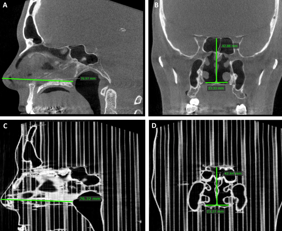
The team first performed cone-beam computed tomography (CBCT) scans on five adult patients with chronic nasal congestion. CBCT was chosen because of its relatively low radiation dose compared to conventional CT scans. Use MATLAB to convert the scanned data into a 3D printable format, and then use Slic3r to prepare and slice it. In order to maintain dimensional accuracy where possible, without supporting, all printing is done on Lulzbot Taz 4 with PLA 3D printingFinished on board.
In order to accurately compare the print with the patient’s nose, a CBCT scan was performed on all parts of the PLA.From the perspective of the maxillary sinus volume of the two scan groups, although it is considered to be very close to the actual value range,3D printingThe parts deviate slightly by 1.05 cubic millimeters.
Finally, compare the airflow resistance of the printed part with the airflow resistance of the patient’s nose. This is done by using an instrument called a nasal pressure gauge, which is made specifically for this purpose. Using it on a patient is simple, but the printed part requires a certain DIY skill, so the researchers connected the tube to the back of the printed part and clipped the other end to the nose for scientific research.The resistance values of the two groups were similar, and the research team concluded that what they could not see before3D printingThe nasal passage method shows great promise for clinical application.
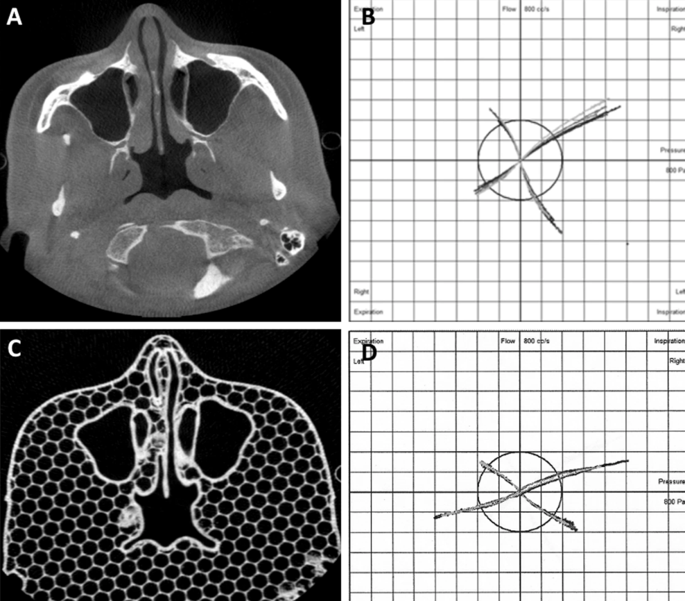
The patient’s CBCT scan and the corresponding PLA nasal pressure measurement model. The picture comes from the University of Tampere.
Medical model3D printingBeyond the academic world. Digital manufacturing service provider Fast Radius and UK-based medical technology company Axial3D recently announced a new “DICOM-to-Print” service for surgeons and hospitals in North America. They plan to improve surgical planning by providing micron-level accurate, patient-specific anatomical models.
Elsewhere in Queensland, researchers published a study that challenges the applicability of FDM in generating anatomical reconstructions. The team claims that errors and defects in the replicas may cause harm to patients through poor treatment plans, and hope that their work will optimize the process.
China3D printingNet compile article!
(Editor in charge: admin)
0 Comments for “Finnish researchers use 3D printing to improve nasal surgery plans”