
In 2020, Liang Ma, Yuting Li and Yutong Wu, etc., published an article titled: The construction of in vitro tumor models based on 3D bioprinting on Bio-Design and Manufacturing.
This review paper focuses on the current
biology
3D printingThe application of technology in the construction of in vitro tumor models.along with3D printingTechnology in Biology
medicine
The popularization and development of the field, the emergence of artificial tissues with complex structures and diverse components, especially in vitro models of heterogeneous multicellular/material tissues, provide reliable molecular biology research for various diseases and the research and development and screening of various drugs. Application platform.This article briefly summarizes the commonBio 3D printingThe strategy and design of bio-inks focused on in vitro tumor models with different structures and their characteristics, and proposed future prospects.
Cancer is characterized by high mortality, complex molecular mechanisms and expensive therapies. The tumor’s microenvironment is composed of a variety of biochemical clues. The interaction between tumor cells, stromal cells and extracellular matrix plays an important role in the occurrence, development, and development of tumors.
Blood vessel
Play a key role in generation, invasion and metastasis. In order to better understand the biological characteristics of tumors and reveal the key factors of cancer treatment methods, the establishment of in vitro tumor models can reproduce all stages of tumor development and simulate tumor behavior in vivo, which is important for achieving efficient and patient-specific drugs Screening and biological research are of great significance. Due to the lack of potential to construct complex structures and angiogenesis, traditional tissue engineering methods for constructing tumor models usually cannot simulate the later stages of tumor development.In the past few decades, biological3D printing technologyIt has gradually been applied in the construction of tumor microenvironment, which can precisely control the composition of tumor-related cells and extracellular matrix components and well-organized spatial distribution.biology3D printing technologyTumor models with multiple scales, complex structures, multiple biomaterials and vascular networks can be established with high resolution and high throughput, and become a multifunctional platform in biomanufacturing and medical research. According to the structure of the model, in vitro tumor models can be divided into the following four types:
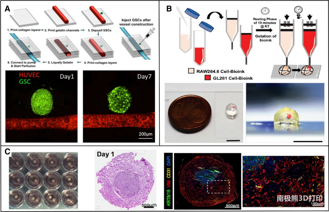
Type 1: In vitro tumor model without scaffold structure
Compared with a flat 2D culture environment, tumors and their stromal cells in a 3D environment can usually show behavioral characteristics and drug responses that are closer to the actual situation in the body. The scaffold-free tumor model composed of various bio-inks and cells can establish a three-dimensional tumor microenvironment, providing a high-throughput experimental model for exploring the interaction between cells and drug screening.
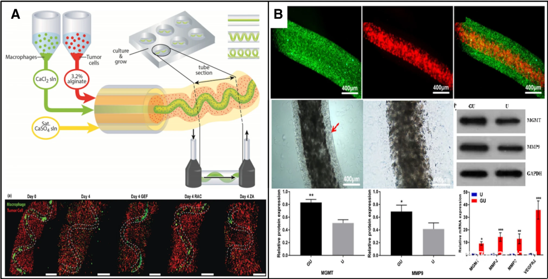
Type 2: In vitro tumor model of fibrous structure
Extrusion typeBio 3D printingConstructed by technology, combined with coaxial printing technology, can carry out the layered space design of the shell-nucleus structure of different types of bio-inks and cells, conduct cell co-cultivation and interaction studies, and can also conveniently construct vascular structures for in vitro tumors The organization provides a more realistic and complex microenvironment model.
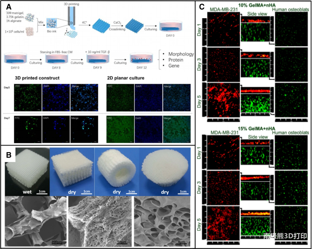
Type 3: In vitro tumor model with mesh scaffold structure
Because tumor models with single-layer and multi-layer mesh scaffold structures are usually easy to construct and have strong versatility, this type of structure is often used in the printing of various bio-inks and cell types.Although the simplified method and function evaluation of the grid structure model
standard
It needs to be further standardized and perfected, but this type of model is more efficient in construction and is conducive to maintaining the activity and phenotype of the cells in the model, and has great potential for application and optimization.
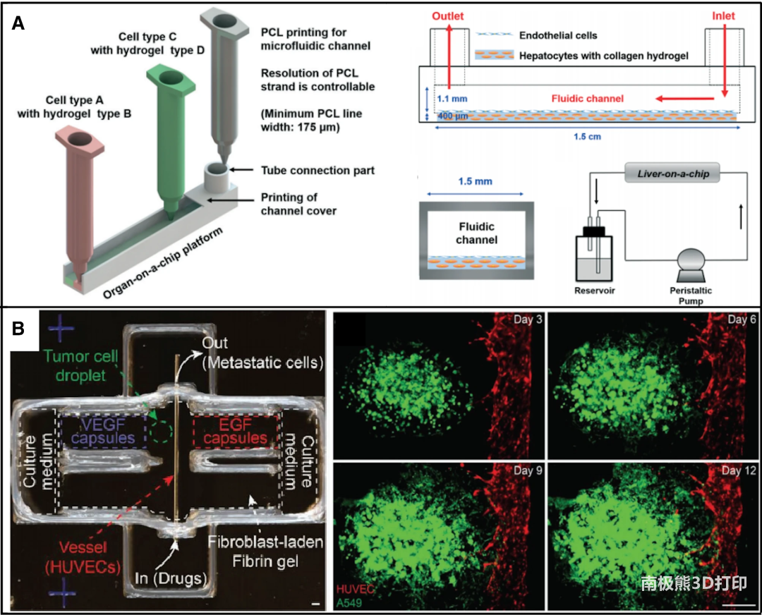
Type 4: Bio-printed in vitro tumor chip
In recent years, with the development of biological 3D printing technology and the advancement of manufacturing processes, the application of organ chips can greatly increase the complexity of in vitro tissue structure models, and the design of tumor chips can have components of tumor microenvironment closer to the real situation in the body And distribution to summarize the various physiological activities in the body and the response to drugs or environmental stimuli, which play a huge value in personalized anti-cancer treatment and drug development.
Prospects of research on in vitro tumor model construction
The improvement of bio-3D printing technology and manufacturing process, as well as the optimization of bio-ink, are of great significance for maintaining the cell viability, phenotype and function of the model. At the same time, the fusion of multi-material and multi-printing strategies also provides the possibility to construct more complex tissue and organ models in vitro. The functional evaluation system of the in vitro biological system and the standardized design of the overall construction process make the further development and application of the in vitro tissue and organ model in the field of biomedicine have broad prospects.
Article Source:
https://doi.org/10.1007/s42242-020-00068-6
(Editor in charge: admin)
0 Comments for “Ma Liang, Zhejiang University, etc.: Construction of an in vitro tumor model based on bio-3D printing”