
|
Introduction: For the first time, researchers from the Massachusetts Institute of Technology (MIT), Northwestern University and other institutionsbiologyIn the case of any polymer other than the material, the biological structure is carried out3D printing. Researchers use gels composed of protein polymer molecules produced by Escherichia coli to design devices that release anti-cancer drugs or capture toxins from the environment.
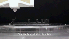
△Microbial ink3D printing
It is reported that the researchers first genetically engineered E. coli cells to produce protein polymer molecules called curly nanofibers. It comes with two modules with opposite charges, called “knobs” and “holes”. As the crimped fibers are produced, they will cross-link and the knob will be locked into the hole. Then, before removing E. coli cells, this material is filtered through a nylon membrane to produce a printable gel that is sufficiently viscous and elastic. This gel is called “microbial ink” by researchers.
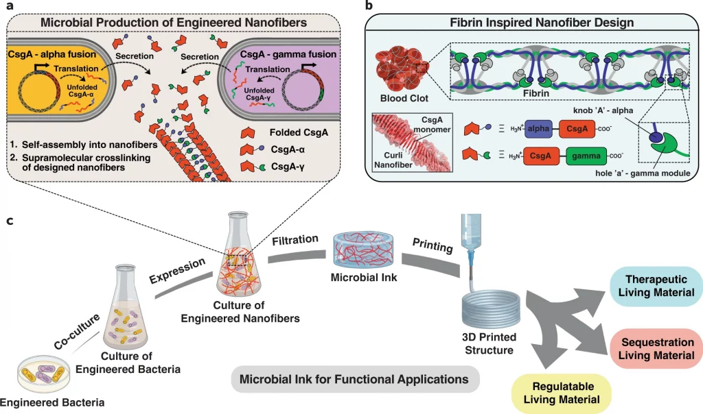
△The α (knob) and γ (pore) protein domains derived from fibrin are fused with CsgA, the main structural component of curled nanofibers, and E. coli is genetically engineered to produce microbial ink. After secretion, CsgA-α and CsgA-γ monomers self-assemble into nanofibers, which are cross-linked through the knob hole binding interaction. The knobs and pore domains are derived from fibrin, and they play a key role in the supramolecular polymerization during the formation of blood clots. Finally, functional biological materials are obtained by printing microbial ink.
Before printing, the bio-ink was sucked into a 10ml Luer-Lok™ syringe and passedstandard27G needle tip, and Fisnar’s prefabricated 1/4 inch blunt end, and the upgraded ANET A8 (Shenzhen Anet Technology Co., Ltd.)3D printingMachine for bioprinting. Through deposition, the researchers were able to produce thin fibers half a millimeter wide. And, they can maintain their strength when stretched between two pillars 16 mm apart.

△Single-layer grid, 10-layer square, 10-layer circular, 21-layer solid cone.Scale bar 1 mm
To further advance the project, the team embedded a new type of E. coli. This Escherichia coli is designed to release the anti-cancer drug azurin. When exposed to a chemical called IPTG, the azure seed structure can release drugs on demand. Researchers have also created various Escherichia coli that can produce crimped fibers that can be combined with bisphenol A (BPA, a toxic substance found in many plastics). After testing, the gel can absorb nearly 30% of toxins from the surrounding liquid within 24 hours.
Related literature: Duraj-Thatte, AM, Manjula-Basavanna, A., Rutledge, J. et al. Programmable microbial ink for 3Dprinting of living materials produced from genetically engineered proteinnanofibers. Nat Commun 12, 6600 (2021).
Document address:https://www.nature.com/articles/s41467-021-26791-x
|
(Editor in charge: admin)
0 Comments for ““Nature Communication” 3D printing microbial ink, can be used for genetic engineering protein nanofiber production”