
China3D printingThe study of cancer biology is a top priority for researchers around the world. From consortia to universities, pharmaceutical companies, newcomers to the drug development industry and research institutions, current research to understand how tumors develop is critical to fighting disease.At the University of Caen-Normandy (UNICAEN) in France, two groups of more than 30 researchers, clinicians and PhD students are developing a new3D bioprintingA tumor model that will provide a novel alternative tool for studying tumor biology and anticancer response to therapy.
Many of these are part of CERVOxy, one of the scientific teams in the Brain and Tumor Pathology Imaging and Therapeutic Strategies (ISTCT) department, which was created in early 2012 by the French National Centre for Scientific Research (CNRS), the Atomic Energy Commission for Energy and Alternative Energies (CEA) and UNICAEN, and hosted on the GIP CYCERON imaging platform in Caen, France. CERVOxy’s scientific team focuses on hypoxia and its role in glioblastoma, a fast-growing brain tumor, and brain metastases.
All of these themes unfold on different axes to study tumorigenic or tumorigenic processes to develop new therapeutic strategies. For example, they are studying how to use hadron therapy (protons and carbon ions) to treat brain tumors. In addition, in vitro and in vivo methods are being used to evaluate the effects of these therapies on healthy brain tissue, which is why they are starting to develop new models based on bioprinting technology.
Nolwenn Pasquet, a postdoctoral researcher at the University of Caen and one of CERVoxy’s researchers, said: “His research focuses on the effects of radiation therapy and hadron therapy on healthy brain tissue in the context of glioblastoma. Pasquet, along with her colleagues, is using Cellink’s INKREDIBLE+ for a lot of work.
“Despite recent improvements, the treatment of glioblastoma remains challenging, and the physiopathology of these tumors is so complex that the use of two-dimensional in vitro models cannot recapitulate the in vivo situation. Furthermore, there is a lack of tools for modeling cellular Relevant models of interactions between, for example, 2D models cannot reflect the tumor microenvironment, such as low oxygen gradients and the presence of surrounding brain cells and inflammatory cells. In this case, new 3D brain models obtained by bioprinting Very attractive for glioblastoma research.”
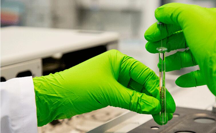
For this study, Pasquet and his researchers used mouse glioblastoma cell lines to develop novel3D bioprintingglioblastoma model. These cells were then embedded in Cellink’s specific bioinks to mimic the extracellular matrix, and the models were then bioprinted with the INKREDIBLE+ bioprinter provided to CERVOxy by the LARIA team at the Franiais Jacob Institute for Biological Studies.

According to Pasquet, in further experiments, by combining glioblastoma cells with surrounding cells (astrocytes, inflammatory cells, etc.) and analyzing their cellular progression, invasiveness, and interaction, crosstalk between glioblastoma cells and surrounding cells (astrocytes, inflammatory cells, etc.) can be observed.“In terms of preliminary results, we observed after bioprinting that the glioblastoma cells had a uniform distribution until six days, and then started to form clusters of cells around the periphery of the models at 14 days of cell culture. Interestingly, these models recapitulates one of the most important features of glioblastoma: hypoxia. Indeed, 14 days after biobromination, we observed a hypoxic gradient in the model with hypoxic cells at the core of the model at the periphery or No observations for six days.”
They also X-rayed the models, Pasquet said. X-ray radiation therapy complements surgery and chemotherapy and is part of the standard regimen for treating brain tumors. Like in medical radiography, it involves delivering photons in varying doses, except in this case to destroy cancer cells.through these3D bioprintingmodel, the researchers wanted to assess the cell’s response and sensitivity to radiation, and thanks to the use of specific markers, they were able to assess the cell’s proliferation, which gave an indication of how the tumor was developing in its surrounding environment.
“Currently, we are starting with this new approach, and it is necessary to further characterize the model and understand its limitations in order to draw conclusions about the results obtained. For example, in this model it is difficult to rule out interactions between cells The fact that it works, real-time microscopy experiments allow us to verify it. This is an important point and part of the reason why we decided to develop this type of model to recreate the microenvironment of these cells within the patient’s brain tissue. These The results are positive and encourage us to continue research in this direction.”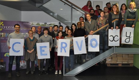
The project is led by the laboratory, a branch of the French National Centre for Scientific Research (CNRS), a publicly funded institution covering all scientific disciplines. It is funded by several sponsors, notably the ARCHADE Centre for Hadron Therapy in Caen; the HABIONOR European project, co-funded by the French Normandy County Council within the framework of the interregional development contract “Vallée de la Seine”, and Région Normandie, for The Normandy Oncology Therapeutic Innovation Network (ONCOTHERA) project.
Pasquet suggested that the information obtained would not be the same without bioprinting. She explained that “the technology is in full development” and that “we’ve only been using it for a short period of time, just over a year, and depending on what you want to learn, there’s an important characterization step to do. test.”
China3D printingNet Comment: It is believed that the models used in bioprinting should be differentiated accordingly, and many models will be used to replicate fully functional organs in the fields of medicine and tissue engineering. 3D bioprintingIt is this aspect that is of particular interest to us in order to provide a new, more complex model for our research.
China3D printingNet original article!
(responsible editor: admin)
0 Comments for “New bioprinted tumor model helps France study biology”