China3D printingAn international team of scientists led by the Universities of Adelaide and Stuttgart has used theminiature3D printingTechnology developed the Optical Coherence Tomography (OCT) endoscope.
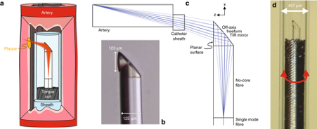
The research team’s novel probe fabrication technique uses laterally free-form micro-optics (less than 130 μm in diameter) directly on single-mode fibers3D printing. The final microscopic imaging device measures only 0.48mm, small enough to access blood vessels and overcome the resolution issues encountered with existing techniques. The improved 3D images provided by enhanced endoscopic cameras could allow doctors to better understand the causes of heart disease, potentially preventing heart attacks before they occur.
Dr. Simon Siller, head of the Optical Design and Simulation Group at the University of Stuttgart, said: “Until now, we have not been able to make such a small, high-quality endoscope so small. Usingminiature3D printing, we can print complex lenses that are too small to be seen by the naked eye. The entire endoscope, with its protective plastic casing, is less than half a millimeter in diameter.
It’s been a pleasure to work on as we take these innovations and build them into such a useful project. It’s amazing what we can do when we put engineers and clinicians together. “

3D printing
The diameter of the device (pictured) is only 0.48mm” alt=” Researcher’s Micro
3D printing
The diameter of the device (pictured) is only 0.48mm” width=”620″ height=”253″ />
Researcher’s Micro3D printingThe device (pictured) is only 0.48mm in diameter. Image from the journal Light: Science and Applications.
3D printing, Endoscopy and Vascular Health
For medical personnel, fiberoptic endoscopes have quickly become an important clinical tool, providing real-time guidance during medical interventions. In particular, OCT endoscopy has been used in a large number of surgical cases, with an estimated 410,000 procedures assisted to date in Australia alone.
Nonetheless, despite its widespread use, endoscopic technology has not been perfected. Miniaturized, high-resolution probes are still in high demand, not only for imaging small, narrow luminal organs, but also for minimizing the discomfort of probe insertion during physician procedures. Often, animals such as mice are also used as models of human disease, and in order to get the most out of such experiments, smaller solutions are required.
Previous research has also found that conventional probes cannot capture images of any structures deeper than 100 μm, limiting their potentially life-saving applications in cardiac care. The main cause of heart disease is plaque, which is made up of fat, cholesterol and other substances that build up on the walls of blood vessels. ‘ explained co-author and lecturer Jiawen Li from the University of Adelaide.
Preclinical and clinical diagnosis increasingly relies on visualizing vascular structures to better understand disease. Tiny endoscopes act like tiny cameras, allowing doctors to see how these plaques form and explore new ways to treat them. “
According to the researchers, current probe fabrication techniques are also inadequate because endoscopes often suffer from spherical aberration, low resolution, or shallow depth of field. Also, while resolution and depth of focus are often trade-offs in existing probes, in small devices their physical apertures are very small and no suitable compromise exists. Furthermore, in OCT imaging, the intravascular probe is deployed within a transparent catheter sheath to protect the patient from trauma during camera rotation.
Optically, the cable cover can cause astigmatism in the camera, causing the camera to lose focus, and current manufacturing methods cannot mitigate this. Although previous methods have focused on splicing existing fiber lenses, these methods also cannot achieve the same resolution as conventional OCT imaging.To overcome these limitations, the research team set out to use two-photon polymerization technology to directly combine micro-optics with a diameter of 125 μm3D printingon monofilament compounds.

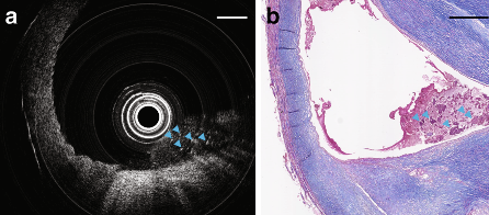
Images captured using the team’s endoscopic device showed necrotic cores of dead cells (pictured). Image from the journal Light: Science and Applications.
Create and test3D printingendoscope
To make their add-on endoscope, the researchers spliced a 450-μm length of coreless fiber into a 20-μcm-long single-mode fiber, expanding the beam before it reached the micro-optical camera.The beam-shaping micro-optics were then directly3D printingto the far end of the material.The researchers’ production method was found to compensate for astigmatism due to the mandatory transparent catheter sheath. Additionally, securing the fiber optic assembly within a thin-walled torque coil allows precise manipulation of the device to the other end of the imaging probe.
To evaluate the performance of their ultra-thin OCT probe for scanning tissue samples, the research team attempted to capture images inside freshly removed human carotid arteries. Although the blocked vessel showed severe stenosis, the team’s ultrathin probe was able to pass through the vessel effortlessly and then be pulled back. In addition, the endoscopic camera was able to detect necrotic cores of dead cells, a key feature in identifying high-risk plaques that can lead to a heart attack.
Further OCT imaging tests were performed on the thoracic aorta of mice. The probe was able to capture images without any noticeable rotational distortion and successfully imaged 3D in very small arteries, the smallest being less than 0.5mm. The picture also shows thick, densely packed fat cells, 15 in diameter


0 Comments for “Researchers develop 3D-printed endoscopic imaging device to improve cardiac care”