China3D printingNet February 27th, scientists at the Wake Forest Regenerative Medicine Research Institute (WFIRM) in North Carolina have discovered a method that can facilitate their development3D bioprintingTechnology to engineer skeletal muscle as a potential therapy to replace diseased or damaged muscle tissue.
Skeletal muscles are responsible for moving the body. Therefore, when they are damaged due to blunt weapon injuries, combat injuries or burns, they do not regenerate, but require reconstruction surgery, and usually connective tissue or adipose tissue is replaced with muscle grafts, and the latter does not have contraction. ability. Over time, the muscles atrophy and these muscle cells die. Although various attempts have been made to restore muscles, such as cell therapy and the use of various biological materials, none of them can promote skeletal muscle repair and functional regeneration. According to WFIRM research, researchers know that effective neural integration of bioengineered skeletal muscle tissue has always been a challenge, but now they bring new hope to patients.A group of researchers in their recent publication entitled “Integrating Nerve Cells intobiology3D printingA study published in the paper “Promoting the Recovery of Muscle Function in the Structure of Skeletal Muscles” studied the effect of integrating nerve cells into bioprinted skeletal muscle constructs to accelerate the regeneration of functional muscles in the body. In other words, they applied the bioprinted construct to a rat model of tibial anterior (TA) muscle defect and evaluated the functional results of muscle tissue reconstruction and innervation.
WFIRM lecturer Jim Ji Hyun Kim said: “This represents a major advancement in the treatment of this type of injury. Our hope is to develop a treatment to help heal injured patients and restore them to as many functions and normal conditions as possible. .”
WFIRM acknowledges that the Institute’s scientists have previously demonstrated that the integrated tissue and organ printing system (ITOP) developed in-house over a 14-year period can produce organized muscle tissue that is strong enough to maintain its structural characteristics. Since then, researchers have been developing and testing different types of skeletal muscle tissue constructs to find the right combination of cells and materials to achieve functional muscle tissue. In the current study, they studied the integration of nerve cells into bioprinted muscle structures to accelerate functional muscle regeneration.
In this particular study, muscle defect injuries in rodents were caused by removing 40% of TA muscles and other injuries that caused irreversible anatomical and functional deformities six months after the injury. After the TA muscle defect was created, they implanted the bioprinted skeletal muscle structure and muscle progenitor cells (MPC) into the defect site for anatomical and functional skeletal muscle regeneration.
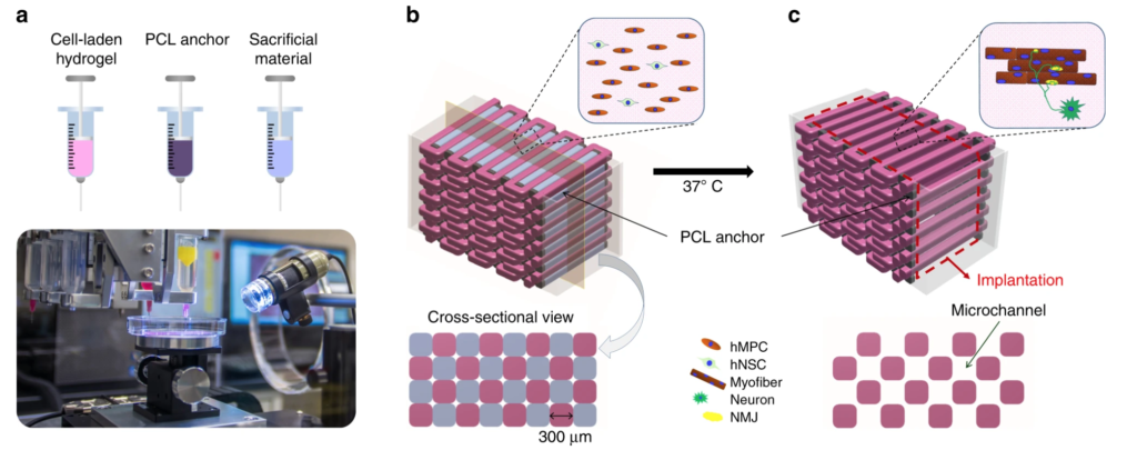
The team used its own custom bioprinting system to create skeletal muscle structures. They described the system as consisting of four distribution modules (including cooling and heating units), air pressure controller, XYZ table and controller, temperature controller and humidifier in a closed room. They used the cell-loaded hydrogel, cell-free sacrificial hydrogel, and supporting polymer for the 3D skeletal muscle structure by loading the bio-ink into different sterile syringes.

3D bioprinting
The system prints three components layer by layer “alt=” bioprinting of human skeletal muscle structure: use WFIRM’s customization
3D bioprinting
The system prints three components layer by layer” width=”620″ height=”245″ />
Bioprinting of human skeletal muscle structure: Customization using WFIRM3D bioprintingThe system prints three components layer by layer (picture: WFIRM)
The results of the study showed that the neural input to the bioprinted skeletal muscle construct showed improvements in muscle fiber formation (multinucleated single muscle cells), long-term survival, and in vitro neuromuscular connection formation. In addition, they claim that bioimprinting constructs with nerve cell integration function help to rapidly innervate and mature into organized muscle tissue, which can restore normal muscle weight and function in rodent models.They also suggested that3D bioprintingThe rapid integration of human neuroskeletal and muscle structures with the host neural network accelerates the recovery of muscle function.
According to China3D printingNetwork understanding, WFIRM is advancing novel bioprinting research to solve many common problems today, such as severe muscle damage, which is one of the most common problems in orthopedic practice worldwide. Researchers are using bioprinting technology developed on site in their own laboratories to fight the failure to obtain functional recovery, in order to discover new ways to treat patients. With a successful in vivo experiment, we hope that the next step will enable the team to further provide solutions for many patients with this disease.
China3D printingnetworkCompiled from: 3dprint.com
(Editor in charge: admin)


0 Comments for “Researchers perform bio-3D printing of rat’s nerve cells to integrate skeletal muscle structure”