China3D printingNet October 27th, researchers from Arizona State University and the University of Southern California have developed3D printingThe microneedle patch can be used to deliver drugs painlessly.Inspired by the hierarchical structure of limpets (aquatic snails with extremely powerful teeth), the team created an enhanced microneedle array that showed increased resistance to long-term use.In addition, the use of magnetic field assist3D printing(MF-3DP) process, the shape of the device can be optimized in the future to provide drugs without making patients feel uncomfortable in clinical trials.

The joint research team’s bioinspired needle is made using iron-infused polymer. The picture comes from the “Advanced Functional Materials” journal.
injection:3D printingEliminate the pain
Hypodermic needles may have been used for more than 150 years due to their low cost and relatively large capacity, but they are usually painful when inserted and generate large amounts of medical waste. In order to solve these problems, the microneedle patch was introduced in the 1970s. This patch is more convenient, can carry a variety of drugs and relieve pain during use.
If you want to perform these microneedles3D printing, You can use custom geometric shapes to create them to improve the effectiveness of different drugs, but so far, this situation cannot be avoided due to lack of accuracy. For example, Fused Deposition Modeling (FDM) and inkjet printing-based methods require extensive and expensive post-processing to enhance and perfect the functions of the equipment.
In addition, previous experimental needles had to be relatively large to provide sufficient strength, but it was found that this increased size would increase the pain during insertion. To overcome these limitations, scientists turned to an unlikely source: limpets. Recent studies have shown that the teeth of marine organisms are composed of many layers of nanofibers and are one of the strongest materials found in nature.According to China3D printingNetwork understanding,
Based on the evolutionary advantages of limpets, the team tried to design a bioinspired needle array with improved mechanical properties and the potential for painless drug delivery.
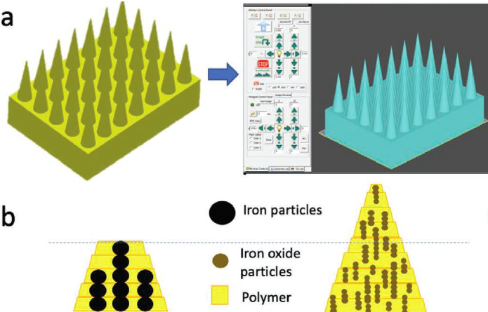
 3D printingCraft to create their microneedles” alt=” The researchers used an innovative magnetic field assist3D printingCraft to create their microneedles” width=”620″ height=”416″ />
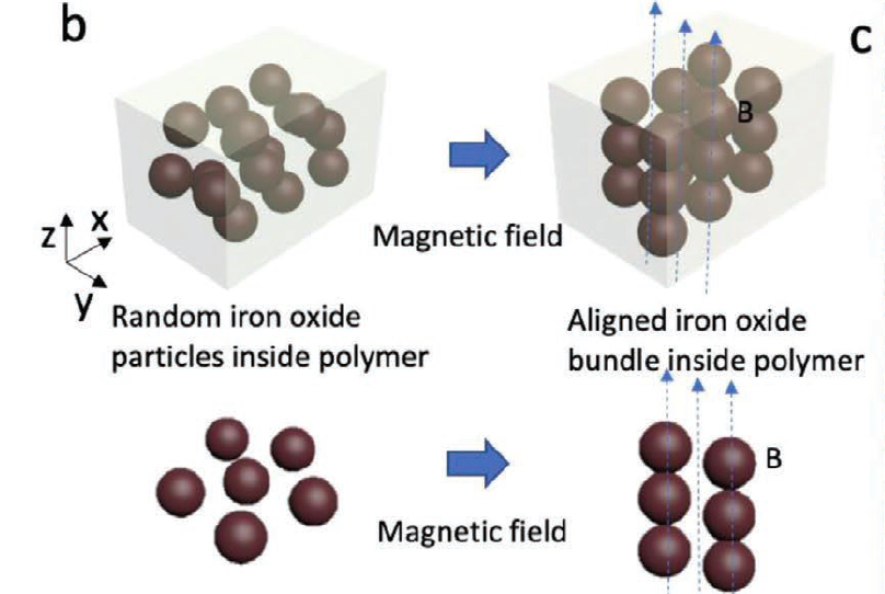
The researchers used an innovative magnetic field assist3D printingCraft to create their microneedles. The picture comes from the “Advanced Functional Materials” journal.
Researchers’ bioinspired microneedle array
The strength of limpet teeth is attributed to the unique arrangement of goethite minerals, making it difficult to replicate using traditional microfabrication methods. As a result, the joint team chose to deploy MF-3DP technology, through which a magnetic field is used to align iron oxide nanoparticles (aIOs) in photocurable polymer materials.
The team then used a stereolithography (SLA) system to selectively cure the composite material and adjust the needle diameter by adjusting the concentration of magnetic particles at different points. The final microneedles were fabricated in a quadrilateral pattern, and the diameter of each conical device was 200 µm, but it was found that the resolution was affected by the width of the needle.
By adjusting the light penetration depth of the printer, the team found that they were able to adjust the width of their device more precisely, resulting in a width of only 8 µm. More importantly, the scientists’ bioinspired array proved to be stronger during testing than the same design printed with pure polymer, which has a lower degree of cross-linking and bends when inserted.
In order to evaluate the pain-reducing elements of its microneedle patch, the researchers applied it to mice and observed no difference in behavior with and without the patch. The team also tested the drug delivery capabilities of its device on pig skin and found that fluorescein can be successfully injected and released within two days.
Overall, the scientists believe that their method is successful because their microneedles exhibit higher mechanical integrity due to the alignment. In the future, the accuracy of the team’s MF-3DP process will help develop needles with customizable micro-features for biomedical and clinical applications.
3D printingDrug delivery system
Microneedle patches have been available since the 1970s, so in recent years3D printingIt is not surprising that advances in technology have led to the development of numerous additive variants.
A team at Rutgers University has deployed a projection micro-stereolithography (PµSL) technology to create bioinspired procedural monoclonal antibody microneedles. Based on the micro-hooks of parasites, the thorny spines of bees and the feathers of porcupines, the device is designed to be horizontally deformable so that it is minimally invasive during insertion.
Similarly, scientists at Temple University also drew inspiration from bees to optimize the design of surgical needles.The group is based on polymer3D printingThe device has a “barb”-like bee-like layout, which they believe can reduce tissue damage during needle insertion.Elsewhere, researchers at the University of Texas at Dallas (UT Dallas) have created a new low-cost method of manufacturing microneedle arrays.By placing the desktop3D printingCombining mechanical and chemical etching technology, the team was able to create fine needles that can be used in multiple medical devices.
China3D printingNet original article!
(Editor in charge: admin)


0 Comments for “Scientists have designed 3D printed microneedle patches for painless drug delivery”