China3D printingNet, September 10, the treatment of cancer has a history of hundreds of years. Researchers are learning how to prevent, diagnose, and treat the world’s biggest killer, and have made significant progress in more complex details. From successful immunotherapy to the growing role of precision medicine, these major advances are helping clinicians save lives. However, there are approximately 14 million new cases each year and nearly 10 million deaths. It is hard to say that the fight against cancer is close to victory. In addition, experts believe that once cancer spreads or metastasizes from one part of the human body to another, the chance of survival is reduced to 10%, which makes it the most deadly feature of the disease.
So far, the management of systemic metastasis has been lacking, but scientists at Lawrence Livermore National Laboratory (LLNL) have developed a unique method that they believe will help clinicians and researchers predict cancer Transmission among individual patients.By puttingbiology3D printingCombining technology with advanced computational process simulation, an expert group believes that they lay the foundation for the development of predictive capabilities to understand the attachment of tumor cells to blood vessels, which is the first step in the formation of secondary tumors in the process of cancer metastasis.
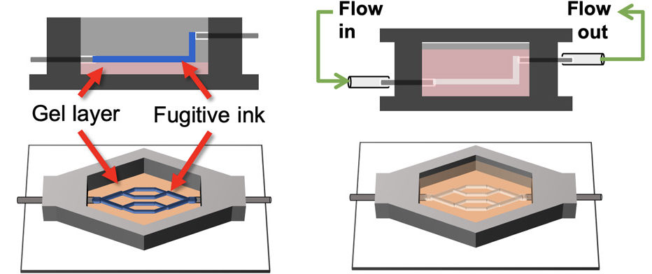
The discovery is described in a paper published in the journal Science Advances. It is a new method of training computational models for biological processes and provides information on how and why cancer cells metastasize in certain areas of the vasculature. Insights. LLNL researchers used a custom extrusion-based bioprinter to print the human brain’s living vasculature in 3D, and paired it with advanced computational simulations to solve the physical problems involved in the spread of cancer through metastasis.

The sacrificial ink method is used to bioprint endothelialized vascular beds with complex geometries. (Photo courtesy of Claire Robertson/LLNL)
According to LLNL biomedical engineer Monica Moya, the lead investigator of the study and lead investigator of LLNL’s bioprinted vasculature device, tumor cells tend to escape from the primary tumor and pass through the vasculature, eventually they attach to blood vessels On the wall, and through the endothelium, it enters the tissue and grows like a seed in the soil, usually in areas such as forks in blood vessels. However, by combining bioengineering and computational methods to analyze the behavior of circulating tumor cells (CTC) and the physical principles of cell binding with vascular endothelial cells (the cell layer located on the inner surface of blood vessels), the team provided new insights into the basic CTC flow Kinetics and adhesion behavior.
“Computational modeling is definitely a useful tool, but you still need to benchmark it with the actual situation.” Moya said, “With this method, we can make biology simple and clean according to the needs of validating models. , And can increase the complexity of biology and computational models. Physics is very important in biology. This article does provide you with a framework for how to use these in vitro models and simulations to sort out the contributions of biology and physics, and indeed Provides strength for the lack of power in this field.”
According to China3D printingWang understands that scientists usually use animal models to understand how physics promotes cancer metastasis, but these models are not entirely useful or have nothing to do with human biology.On the contrary, her team has used human brain endothelial cells3D printingThe vasculature and keep it flowing in the fluid platform, creating an extracorporeal system.
After the cells completely covered the channel of the device, they were aligned in the blood vessel. About a week later, the researchers injected breast cancer cell lines into the device to observe how and where tumor cells began to metastasize in the newly formed cerebral vascular system. Experts believe that certain areas of the brain are more prone to breast cancer metastasis, but the mechanism of local sensitivity is still poorly understood and cannot be prevented. This is why this research is essential to understand the behavior of breast cancer cells once they metastasize.

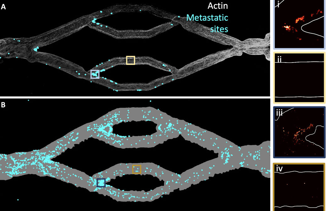
The attachment site of CTC shows the difference between decellularization (B) and endothelialization (A) channels. Compared with endothelialization, the intact acellular vascular bed exhibits a greater burden of CTC. (Photo courtesy of Claire Robertson/LLNL)
The researchers said that after the tumor cells circulate at a physiological flow rate, Lawrence researcher Claire Robertson (Claire Robertson) is committed to the development of an early breast cancer model. Biophysics was compared. Then, these experimental results are compared with the 3D calculation simulation that replicates the geometry collected from the 3D map to reproduce the precise geometry of the bioprinted blood vessel, which enables high-precision fluid dynamic analysis of the attachment conditions.
The lead author and LLNL researcher engineer said: “Using this advanced bioprinting method to engineer functional, instillable human cerebrovascular systems is extremely challenging, but we now have a strong control over the technology, and It is possible to create a variety of living human tissue structures. Using this method, we can test, observe, and measure biological phenomena that were impossible before. We will continue to iterate on these findings to clarify how circulating tumor cells are and When to choose its target in vivo. By pairing our engineering platform with a computational model, we can directly query the behavior of metastatic cells and the rules that control metastatic cells, which is much faster than conducting experiments alone.”
China3D printingNet Comments: This new method allows researchers to separate the biological and physical roles that drive organ metastasis and colonization. The researchers expect that the simulation will be used to predict the location of tumor spread, so as to screen high-risk patients and target the most vulnerable areas for therapeutic intervention. According to Moya, this can provide clinicians with another way to treat patients, allowing them to use MRI to simulate the location where circulating tumor cells may get stuck, so that they can concentrate on greatly improving the effectiveness of the treatment.
China3D printingnetCompile the article!
(Editor in charge: admin)


0 Comments for “Scientists use biological 3D printing and computer modeling to predict cancer growth”