China3D printingNet, March 15th, Slovak researchers are further studying the use of PLA in bioprinting, and in the recently published “Used in Bone Tissue Engineering”3D printingPublished their findings in “Porous Polylactic Acid-based Scaffolds: An In Vitro Study”.
The team created samples in the form of PLA scaffolds, evaluated cytotoxicity and biocompatibility, and its ultimate goal is tissue engineering of bone tissue to assist bone regeneration (one of the most challenging areas). Researchers have conducted a wide range of different studies on bioprinting and bone regeneration, creating various structures, scaffolds, and using different printing methods.
PLA has usually been used before, and the researchers here used commercially available PLA scaffolds. They divided the samples into three different groups, each of which was pre-processed as follows:
.
Complete medium
.Bovine Fetal Serum
.Human blood
In addition, the research team analyzed periosteal-derived cells in terms of cytotoxicity and biocompatibility.

The nature of waste materials and the nature of the printing process
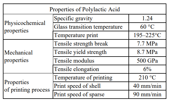
The bracket is using FFF 3D printingCreated, the scaffold sample is 10*10*4 mm, and the porosity is 61%. The periosteum was taken from the proximal tibia of a 55-year-old female patient who was undergoing knee replacement surgery. With her consent, the ethics committee of the Louis Pasteur University Hospital in Kosice approved the procedure.
And inoculate 25,000 cells/cm2 in a 25 cm2 culture flask (T25), which contains Alpha-modified minimal essential medium (Invitrogen, GIBCO®, U.S.) and added 10% fetal bovine serum (FBS, Invitrogen) , GIBCO®, USA) and 1% ATB,” the researcher explained.
After 5 days, non-adherent cells were removed by changing the medium. The adherent cells were cultured in a humidified atmosphere of 5% CO2 at 37°C under standard culture conditions, and the medium was changed every 2-3 days. The confluent cell layer was dissociated with 0.05% trypsin-EDTA solution (Invitrogen, USA), and the number and viability of the cells were evaluated by TC10™ automated cell counter (Bio-Rad Laboratories). Periosteum-osteoprogenitor cells (PDO) from the third generation (P3)-are used for flow cytometry analysis and co-culture with the scaffold. “
Sterilize the stents and divide them into three groups:
“The first group was incubated in human serum, the second group was supplemented with 10% FBS and 1% ATB in a medium containing Alpha-modified minimal essential medium, and the third group was incubated in 10% FBS.”

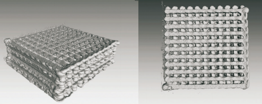
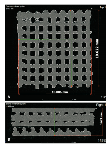
Printed scaffold prepared by FFF technology, displayed by computer tomography

The output of the scaffold-the internal structure of the scaffold:
A) Top view (119%), B) Right view (167%)
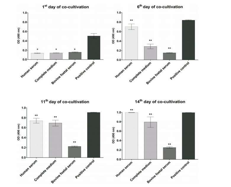
The cells were measured four times and compared with the control group. The result is good biocompatibility. However, the researchers pointed out that it works best in stents coated with human serum. This treatment also promotes cell growth.

During co-cultivation with PLA scaffolds, the proliferation of periosteal-derived osteoprogenitor cells was measured by the CellTiter96® AQueous One Solution cell proliferation assay after 1, 6, 11, and 14 days. The data represents the average ± SD value of four independent measurements. The value of p is p <0.05 on the first day of co-cultivation
and p <0.01 on the other days of co-cultivation (**)During the two weeks of culture, SEM was also used to observe the distribution, adhesion and proliferation of human PDO on the natural PLA scaffold. Human PDO showed good viability in the scaffold. It was incubated in human serum for 48 hours, which was expressed by enhanced cell proliferation and proliferation, and the pH of the medium in which the scaffold and cells were co-cultured was 7.4 after 14 days. . The researchers concluded: “The pH has reached the pH of human blood.
The obtained PLA porous scaffold is beneficial to the periosteum attachment to progenitor cells and proliferation. In addition, the cells penetrate the scaffold through the interstitial pores, which is of great significance for cell compatibility evaluation. “China3D printingNet Comments: New strategies, such as the use of scaffolds, combined therapy with healing promoting factors and stem cells, and finally3D printingTechnology
Although it is still in its preliminary stages, it may provide new and exciting alternatives in the near future.
(Editor in charge: admin)


0 Comments for “Slovakia tries 3D printed PLA scaffold for bone regeneration”