
Blood vessel
It is found in almost all tissues of the human body and has an important impact on human pathophysiology.However, at present, for multiscale vascular
biology
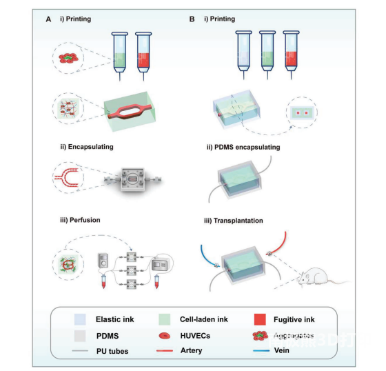
Manufacturing still faces a lot of problems. Researcher Gu Qi from the Institute of Zoology, Chinese Academy of Sciences, together with researcher Zheng Xiongfei, Shenyang Institute of Automation, Chinese Academy of Sciences, and Wang Shu Research Institute, Institute of Chemistry, Chinese Academy of Sciences, developed a multi-material printing method for vascularized tissue using a customized printer. And there is a huge application prospect in liver tissue engineering. The related paper “3D Liver Tissue Model with Branched Vascular Networks by Multimaterial Bioprinting” was published in the journal Advanced Healthcare Materials.
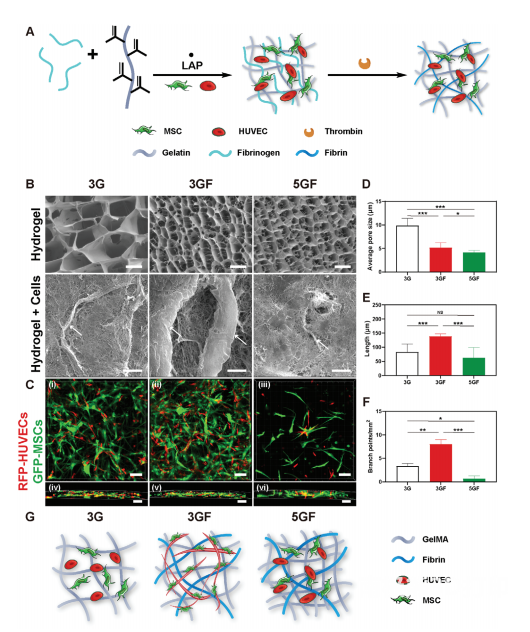
To combine the optimal printing properties of GelMA with the angiogenic properties of fibrin, the researchers used a cell-loaded ink consisting of a mixture of GelMA and fibrin. SEM images showed that the average size of connected pores increased with decreasing GelMA concentration and decreased after fibrin addition. The porous structure facilitates the diffusion of nutrients and improves cell survival. To assess the biocompatibility of the inks, cells containing different fluorophores were co-encapsulated in GF matrices and confocal microscopy was used to quantify the extent of capillary-like network formation. The results showed that capillary-like structures formed in the hydrogel. Capillary-like structures formed in 3% GelMA + 0.25% fibrin (3GF) were more abundant and longer than in 3% GelMA (3G) or 5% GelMA + 0.25% fibrin (5GF). Therefore, in the following study, 3GF was selected as the cell-loading ink. (figure 2)
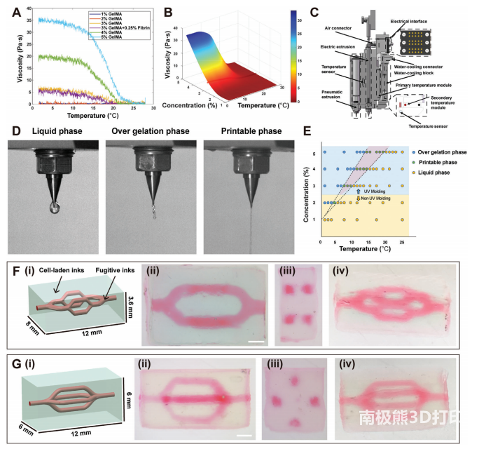
The viscosity of the ink is one of the important factors affecting the printing process. To this end, the researchers evaluated the viscosity of GelMA at different temperatures. And by fitting the viscosity data, a three-dimensional heat map representing viscosity-temperature-concentration was established. It was found that 3% GelMA can achieve the lowest concentration with controllable viscosity at temperatures above zero. GelMA was extruded using an electric drive, and the temperature control system was adjusted to keep the bioink in a gel state. To design and fabricate vascularized tissue structures, the researchers designed and printed several different structures to demonstrate the formation of stable printed structures with channel structures. (image 3)
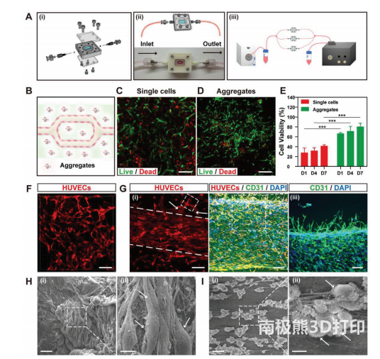
To provide stable perfusion for long-term cultures, the researchers fabricated a continuous flow perfusion system. HUVECs and MSCs were printed as single cells coated with 3GF and compared to study the viability of single cells and multiple cell aggregates during perfusion culture. It was found that the printed cells could proliferate over time. Next, to demonstrate the formation of vascularized tissue, the researchers used HUVEC and HFF co-culture aggregates to print, and after 3 days of culture with 3GF hydrogels, morphological assessments of vascularization and lumen formation were performed. It was found that the printed tissue showed multiple cell aggregates and formed a capillary-like network. HUVECs were implanted into the constructed channels, and after overnight continuous perfusion culture, the cells aggregated and a high cell density could still be maintained. (Figure 4)
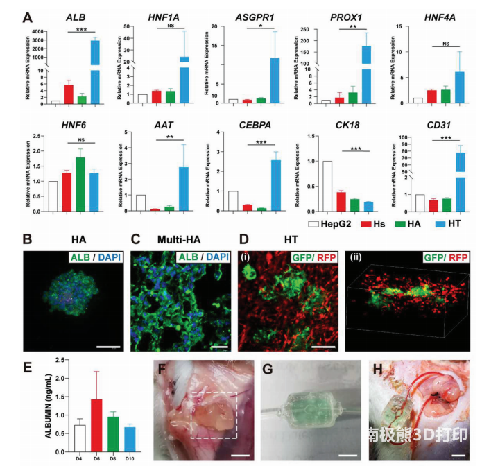
To construct a liver model in vitro, the researchers encapsulated HUVECs, MSCs, and HepG2 aggregates in hydrogels to observe the distribution of cells and the expression of related genes. The printed tissue showed that HUVECs and MSCs formed a capillary-like network with multiple cell aggregates. The function of the liver unit was assessed by measuring the expression of relevant genes by PCR. Finally, liver tissue cultured for 7 days was selected for further experiments. The printed tissues were implanted subperitoneally in mice, and 3GF hydrogel was chosen as a control. As a result, it was found that new blood vessels appeared in the liver tissue on the 7th day after transplantation.Finally, the researchers demonstrated that these tissues can be directly used in rat arterial vascular transplantation.
Operation
Anastomosis of host blood vessels. (Figure 5)
Taken together, this study demonstrates for the first time that vascularized tissue can be printed using extrusion-based printing using low concentrations of GelMA and fibrin as bioinks.This is based on3D printingOur method allows us to construct vascular networks with active tissue function in centimeter-scale tissue. In addition, it opens up a feasible avenue for drug screening, the study of human tissue development and disease, and the identification of liver tissue-engineered transplantation therapies.
references
Xin Liu, Xinhuan Wang, et al. 3D Liver Tissue Model with Branched Vascular Networks by Multimaterial Bioprinting. Advanced Healthcare Materials
https://doi.org/10.1002/adhm.202101405
(responsible editor: admin)
0 Comments for “Three-dimensional vascularized liver tissue model based on multi-material printing”