
Introduction: On September 15, 2021, researchers from Umeå University in Sweden introduced a new detection method that allows the study of specific cell types in human organs with micron-level accuracy. This method can be used to reveal previously unrecognizable changes in the pancreas, but it can also be used to study other human organs and diseases.
Molecules from Umeå University
medicine
Professor UlfAhlgren said: “This method may help to understand the relationship between cell changes and different disease conditions.”
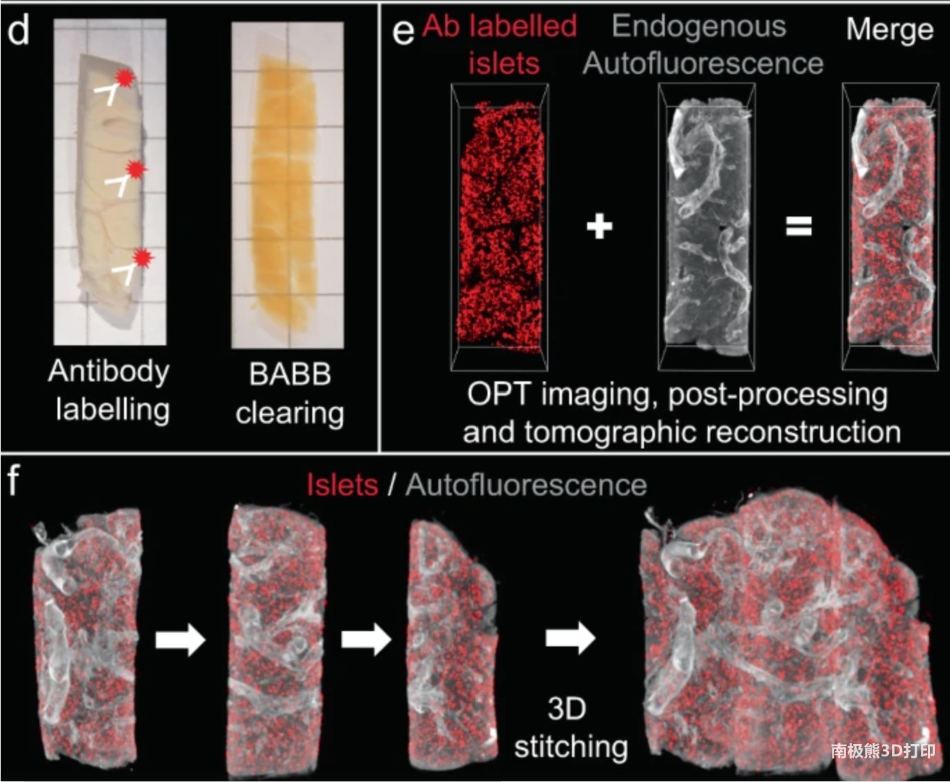
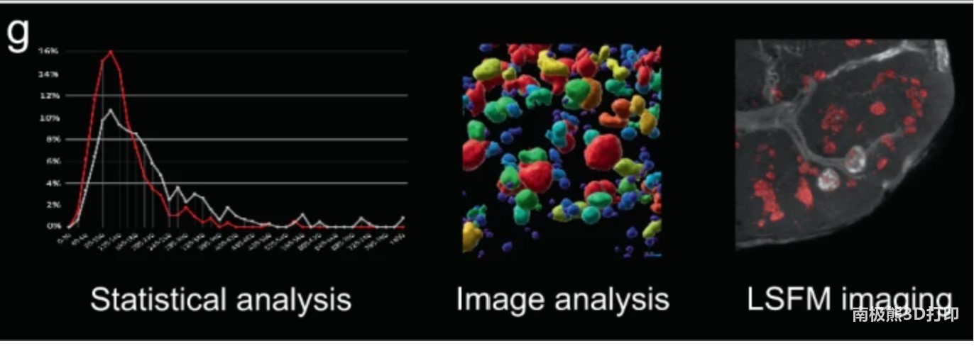
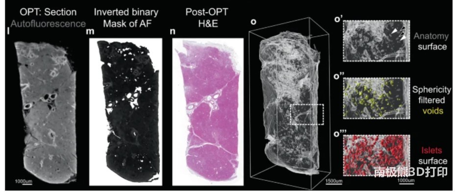
What the researchers did is use3D printingThe matrix is used to segment the organs, and the 3D technology is used to create the most suitable tissue parts for optical imaging. These fragments can then be labeled to visually see any cell type or protein selected. Since each tissue has known coordinates, a single 3D image can be pieced together into a three-dimensional puzzle with a computer to form a complete human organ.
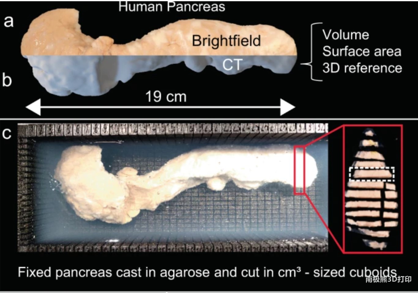
This method allows us to create high-resolution 3D images of human organs of almost any size with micron-level accuracy (smaller than dust particles).Prior to this, it has been possible to create
biology
High-resolution images of materials, and the researchers also used these techniques in this study. On the contrary, the problem is that the previous methods did not provide usable methods to label the various cell types or proteins you want to study, such as using fluorescent antibodies, when you study samples on a larger scale, such as whole organs. This is the problem that the new method has now solved.
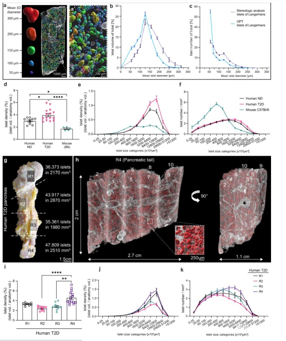

“In addition to using new methods to study diabetes, it can also improve the understanding of other pancreatic diseases, especially pancreatic cancer. We have begun to collaborate with clinical researchers in Umeå to study this.” But the technology itself should be able to be similar The method is used to study other organs and diseases because it can study where cell changes occur in the entire organ environment, their number, and the relationship with nearby tissues and cell types.
(Editor in charge: admin)
0 Comments for “Umeaa, Sweden; University’s new detection method enables 3D microscopic imaging of human organs”