
Bioactive ceramics refer to materials that can promote the growth of new tissues such as bone and cartilage. In addition to containing biologically active materials. When manufacturing bone regeneration tissue engineering scaffolds from such bioceramic materials, it is required that the ceramic materials are relatively hard, can promote cell adhesion, and have osteoinductive, osteoconductivity and/or chondrogenic activity.
Bioactive ceramic materials support bone formation and cartilage formation under specific differentiation medium conditions. Hydroxyapatite (HAp) belongs to this type of bioactive ceramics, which has the mineral composition of natural bones. Hydroxyapatite has osteoconductive and osteoinductive properties, and has the ability to promote new natural bone growth in and around the material.
Composite material of hydroxyapatite (HAp) and polycaprolactone (PCL) or polylactic acid-co-glycolic acid (PLGA)3D printingTissue engineering scaffolds can be formed by hot melt3D printingTechnology for manufacturing. However, there are still many problems in the manufacture of such composite tissue engineering scaffolds.The research team of Dr. Ramille N. Shah from Northwestern University and University of Illinois Health University has developed a method that can be carried out in a relatively low temperature environment.3D printingThe bioactive ceramic ink can be used to manufacture bone regeneration scaffolds implanted in the body. The scaffolds have ultra-high elasticity and the material contains a high proportion of hydroxyapatite.
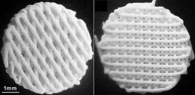
Super-elastic bioactive ceramic bone regeneration scaffold. Source: US20180243484
![]() Facing a variety of tissue engineering applications
Facing a variety of tissue engineering applications
The research team pointed out3D printingThe problems that exist in the manufacture of bone regeneration scaffolds with hot melt printing technology include:
1) Hot melt printing technology does not support the direct incorporation of biologically active factors into the material; 2) The material extrusion process relies on a meltable hot melt suspension. When the weight fraction of solid hydroxyapatite particles is greater than about 0.5, the actual The above cannot be achieved; 3) Because the material is mainly PCL, the hydroxyapatite particles are encapsulated and not exposed on the surface of the material, which means that the surface properties of the material are those of PCL material, namely Smooth and not easy to adhere to cells; 4) In order to prevent deformation or deformation after manufacturing, the final tissue engineering scaffold made of this type of composite material is hard and fragile; 5) Temperature-sensitive materials such as hydrogels or cell materials, It cannot be used as a material for this process.
To overcome these problems, the research team developed an ink formulation containing biologically active particles, and also developed3D printingTissue engineering scaffold and3D printingMethod, and method of cultivating living body tissue on the tissue growth scaffold.
In addition to biocompatibility, the materials used to manufacture tissue engineering scaffolds also require the polymer binders to be easy to process, elastic and biodegradable. The biocompatible polymer binder can also be a bioactive material. Suitable polymer binder materials include biocompatible bioactive polyesters, such as polylactic acid, polyglycolic acid, polylactic acid-co-glycolic acid ( PLGA) (also known as polylactide-co-glycolide (PLG)), polycaprolactone (PCL) or any combination. PCL and PLGA are synthetically derived polyesters that are elastic, degradable, can be used for cartilage tissue regeneration, and have shown the ability to support the differentiation of stem cells into osteogenic and chondrogenic pathways.
The use of elastic polymer adhesives (such as PCL and PLGA) can improve3D printingThe robustness of the bone tissue engineering scaffold.exist3D printingWhen the equipment extrudes ink, the elastomer binder provides continuity, flexibility and firmness, and elasticity to the material. The bone regeneration capacity of the above materials depends on the chemical and mechanical properties of the materials. For example, stem cells will respond to the hardness of the material. In experiments involving HAp and PCL, HAp-induced stem cells mainly follow the osteogenic differentiation pathway, while PCL-induced stem cells mainly follow the chondrogenic differentiation pathway.
Because this ink material can be formulated and processed at a relatively low temperature (for example, room temperature)3D printing, So you can add biologically active factors (such as proteins, peptides, growth factors, and genes) and/or pharmaceutical compounds to the ink, and make it at this temperature3D printingThe structure will not undergo thermally induced degradation. Another advantage of low-temperature processing is that inks with high bioactive ceramic content can be formulated. For example, the content of bioactive ceramics accounts for 60%-90% of the total weight of the ink.In addition, it is allowed to be used with temperature-sensitive materials such as hydrogels and hydrogels containing living cells3D printing.3D printingTechnology has advantages in the field of bone regeneration tissue engineering scaffold manufacturing, because it provides a regular geometric pattern of the layers that make up the scaffold, and the porosity, pore size and pore interconnectivity of the scaffold can be precisely controlled and adjusted.
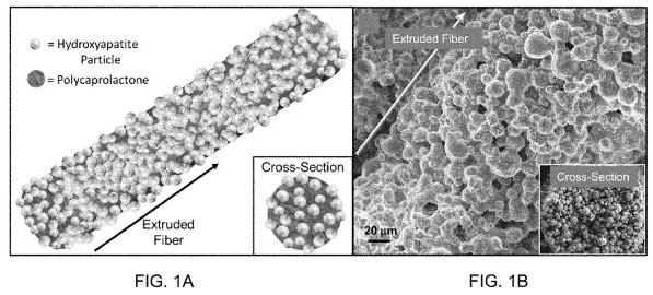
figure 1. Source: US20180243484
The research team is based on the above inks and3D printingThe method was verified. In one of the verification examples, the research team used an ink composed of micro- or nano-sized HAp particles combined with a biocompatible elastic PCL material (Figure 1).
The material used in the example has the following characteristics: rough surface dominated by exposed HAp particles; PCL-dominated macro-mechanical properties (elasticity, large elongation at break); HAp-dominated micro-mechanical properties; biodegradable; osteoinduction ; Complex porous structure.
To ensure that HAPCL materials and printed and post-processed materials are non-toxic and harmless, and to measure the biocompatibility and bone regeneration potential of the materials, the researchers analyzed the3D printingThe material was subjected to in vitro cell research, and HAp scaffolds with 200 μm pores were fabricated from the material.
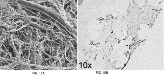
Figure 10. Source: US20180243484
Figure 10 shows the collagen synthesis and deposition of hMSC on the HAPCL scaffold, such as: (A) SEM; (B) histological image. hMSC actively deposits collagen and other extracellular matrix elements in HAPCL.
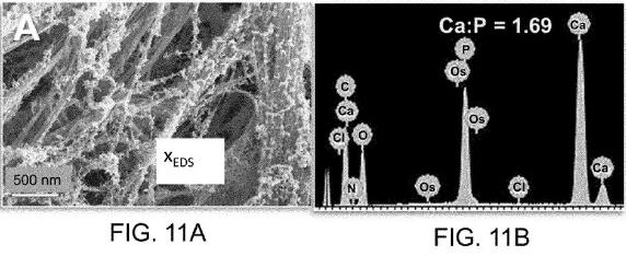
Figure 11. Source: US20180243484
Figure 11 shows the synthesis and deposition of hydroxyapatite by hMSC 28 days after inoculation, such as: (A) SEM; (B) the corresponding energy dispersive X-ray spectra of deposited minerals. hMSC actively synthesizes and deposits hydroxyapatite (calcium-phosphorus ratio 1.69) in HAPCL. The natural hydroxyapatite found in bones is 1.67, and the HAp used in stent manufacturing is 1.59. The HAp produced by the cells in HAPCL is more natural than the original scaffold. These results all indicate that the stem cells on the HAPCL material are viable, proliferate, actively produce extracellular matrix and are undergoing osteogenic differentiation.
The above-mentioned 3D printing bioceramic ink can be used in a variety of tissue engineering applications, including the replacement and regeneration of meniscus, cartilage and subchondral bone (ie for osteoarthritis, cartilage defects and meniscus tissue damage); other cartilage tissues ( (I.e. ear, nose, esophagus, trachea); ligament bone fixation device, used to improve the integration of ligament repair surgery and restore its mechanical function; craniofacial regeneration implants (such as skull plate, nose, bone); corrective surgery Tissue support and regeneration after treatment of pallet cleft plate; support and regeneration of alveolar after tooth extraction; spinal fusion and regeneration; regeneration of any long bones, hip bones or limb bones (ie, hands, wrists, ankles, feet, toes); drugs Or the delivery of growth factors, and biodegradable implants or coatings.
![]() Review
Review
The above research team has conducted animal experiments on super-elastic bioactive ceramic scaffolds. The paper published by the team was published in the May 2019 issue of Plastic and Reconstructive Surgery. The paper reported a3D printingThe super-elastic bone synthetic material, which is expected to become a cost-effective new material for cranio-maxillofacial repair. Preliminary research results show that the material accelerates bone regeneration in rats with skull defects.
The research team stated that there is a large demand for cost-effective bone replacement materials in the field of cranio-maxillofacial reconstruction, and super-elastic bone has great potential to be applied in this field.In animal experiments, in addition to using3D printingIn addition to the repair of skull defects with super-elastic bone, animal autogenous and repair brackets that do not contain bone mineral materials are used to repair other skull defects. Autologous bone is the preferred material for reconstruction of bone defects, but this natural material is difficult to obtain. It is necessary to take bones from other parts of the body, and sometimes it is impossible to obtain suitable autologous bone.
The experimental results show,3D printingThe super elastic bone provides good bone regeneration. In follow-up CT scans, compared with autogenous bone, the effective rate of hyperelastic bone was about 74% after 8 weeks, and the effective rate was 65% at 12 weeks. However, the defect repaired by the scaffold containing only polyglycolic acid material and no bone mineral material showed little new bone formation. Microscopic examination showed that the superelastic bone scaffold was first gradually surrounded by fibrous tissue and then surrounded by new bone cells. Over time, the scaffold will gradually be completely replaced by new bone, combined with the implanted bone mineral.
(Editor in charge: admin)
0 Comments for “Ultra-high elasticity bioceramic bone regeneration scaffold material that can be 3D printed at low temperature”